
Page 161 - Clinical Small Animal Internal Medicine
P. 161
16 Imaging in Cardiovascular Disease 129
(a) Dobermann (b) French Bulldog
VetBooks.ir
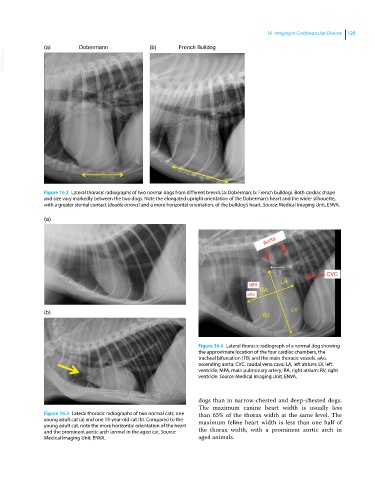
Figure 16.2 Lateral thoracic radiographs of two normal dogs from different breeds (a: Doberman; b: French bulldog). Both cardiac shape
and size vary markedly between the two dogs. Note the elongated upright orientation of the Doberman’s heart and the wider silhouette,
with a greater sternal contact (double arrows) and a more horizontal orientation, of the bulldog’s heart. Source: Medical Imaging Unit, ENVA.
(a)
Aorta
CVC
MPA
aAo
(b)
Figure 16.4 Lateral thoracic radiograph of a normal dog showing
the approximate location of the four cardiac chambers, the
tracheal bifurcation (TB) and the main thoracic vessels. aAo,
ascending aorta; CVC, caudal vena cava; LA, left atrium; LV, left
ventricle; MPA, main pulmonary artery; RA, right atrium; RV, right
ventricle. Source: Medical Imaging Unit, ENVA.
dogs than in narrow‐chested and deep‐chested dogs.
The maximum canine heart width is usually less
Figure 16.3 Lateral thoracic radiographs of two normal cats, one than 65% of the thorax width at the same level. The
young adult cat (a) and one 15‐year‐old cat (b). Compared to the maximum feline heart width is less than one half of
young adult cat, note the more horizontal orientation of the heart
and the prominent aortic arch (arrow) in the aged cat. Source: the thorax width, with a prominent aortic arch in
Medical Imaging Unit, ENVA. aged animals.