
Page 300 - Clinical Small Animal Internal Medicine
P. 300
268 Section 3 Cardiovascular Disease
VetBooks.ir
ARVC
RA, RV dilation
HCM
phenotype
Dietary taurine
deficiency
Increased LV wall thickness Tachycardia-mediated
cardiomyopathy
End-stage
HCM DCM
Systolic phenotype
dysfunction Chamber dilation,
systolic
dysfunction
RCM
phenotype ?Unclassified
Normal LV,
LA dilation Abnormalities not
fitting other
categories
Hypertension
Acromegaly “Transient Hyperthyroidism Anemia
myocardial
thickening”
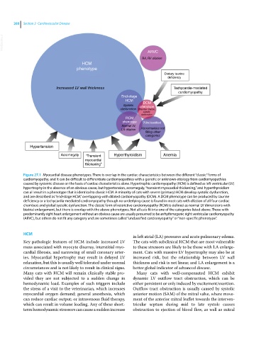
Figure 27.1 Myocardial disease phenotypes. There is overlap in the cardiac characteristics between the different “classic” forms of
cardiomyopathy, and it can be difficult to differentiate cardiomyopathies with a genetic or unknown etiology from cardiomyopathies
caused by systemic disease on the basis of cardiac characteristics alone. Hypertrophic cardiomyopathy (HCM) is defined as left ventricular (LV)
hypertrophy in the absence of an obvious cause, but hypertension, acromegaly, “transient myocardial thickening,” and hyperthyroidism
can all result in a phenotype that is identical to classic HCM. A minority of cats with severe (primary) HCM develop systolic dysfunction,
and are described as “end‐stage HCM,” overlapping with dilated cardiomyopathy (DCM). A DCM phenotype can be produced by taurine
deficiency or a tachycardia‐mediated cardiomyopathy though no underlying cause is found in most cats with dilation of all four cardiac
chambers and global systolic dysfunction. The classic form of restrictive cardiomyopathy (RCM) is defined as normal LV dimensions with
biatrial enlargement, but there is overlap with the above phenotypes. Not all cats fit into one of the categories listed above. Those with
predominantly right heart enlargement without an obvious cause are usually presumed to be arrhythmogenic right ventricular cardiomyopathy
(ARVC), but others do not fit any category and are sometimes called “unclassified cardiomyopathy” or “non-specific phenotype”.
HCM
in left atrial (LA) pressures and acute pulmonary edema.
Key pathologic features of HCM include increased LV The cats with subclinical HCM that are most vulnerable
mass associated with myocyte disarray, interstitial myo- to these stressors are likely to be those with LA enlarge-
cardial fibrosis, and narrowing of small coronary arter- ment. Cats with massive LV hypertrophy may also be at
ies. Myocardial hypertrophy may result in delayed LV increased risk, but the relationship between LV wall
relaxation, but this is usually well tolerated under normal thickness and risk is not linear, and LA enlargement is a
circumstances and is not likely to result in clinical signs. better global indicator of advanced disease.
Many cats with HCM will remain clinically stable pro- Many cats with well‐compensated HCM exhibit
vided they are not subjected to a sudden change in dynamic LV outflow tract obstruction, which can be
hemodynamic load. Examples of such triggers include either persistent or only induced by excitement/exertion.
the stress of a visit to the veterinarian, which increases Outflow tract obstruction is usually caused by systolic
myocardial oxygen demand; general anesthesia, which anterior motion (SAM) of the mitral valve, where move-
can reduce cardiac output; or intravenous fluid therapy, ment of the anterior mitral leaflet towards the interven-
which can result in volume loading. Any of these short‐ tricular septum during mid to late systole causes
term hemodynamic stressors can cause a sudden increase obstruction to ejection of blood flow, as well as mitral