
Page 61 - BSAVA Manual of Canine and Feline Head, Neck and Thoracic Surgery, 2nd Edition
P. 61
BSAVA Manual of Canine and Feline Head, Neck and Thoracic Surgery
include potentially improved cosmesis, a lower risk of sub-
cutaneous emphysema and better exposure to the caudal
VetBooks.ir recommended if exposure of the frontal sinuses is required.
ventral nasal cavity and nasopharynx. This approach is not
The animal is placed in dorsal recumbency with the
man dible taped open or with a mouth gag in place. A midline
incision is made through the oral mucosa and the mucoperi-
osteum of the palatine bone, and the mucoperio steum is
reflected bilaterally. Stay sutures are placed in the mucoperi-
osteum to aid in retraction and improve exposure. The
caudolateral limits of the dissection are the major palatine
arteries, which are identified and preserved. The incision can
be extended caudally to include the soft palate if increased
exposure of the nasopharynx is required. The palatine bone
is removed with an air burr or rongeurs; no effort is made to
save or replace the bone flap. The wound is closed in two
layers using absorbable suture material in a simple inter-
rupted or continuous pattern. A soft, pliable suture material (a)
is used in the oral cavity to minimize lingual irritation.
Postoperative considerations specific for this tech-
nique include a need to protect the mucosal incision
during the healing process. The animal is fed soft food for
1 month and not allowed access to chew toys, bones or
sticks. The incision should be inspected weekly for 2–3
weeks to ensure proper healing. If this procedure is per-
formed on a growing dog, growth deformities are possible
(Holmberg et al., 1989).
Other approaches
An alveolar mucosal rhinotomy approach has been des-
cribed to remove a dorsally displaced canine tooth (Priddy
et al., 2001). This approach is performed through the oral
cavity and provides a narrow window into the most rostral
aspect of the nasal cavity. Limited lateral rhinotomies have
also been described in combination with maxillectomy for
tumour removal. A proper lateral rhinotomy was described
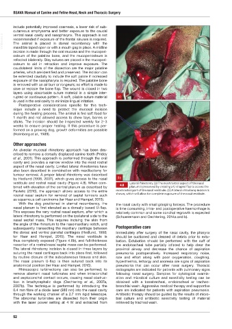
by Hedlund (1998, 2007), which gives access to the nasal (b)
vestibule and rostral nasal cavity (Figure 4.8). When com- Lateral rhinotomy. (a) The most rostral aspect of the nasal
4.8
bined with elevation of the central planum as described by planum is preserved by creating a -shaped flap to access the
Pavletic (2010), the approach allows access to the entire most rostral part of the nasal vestibule. (b) A lateral rhinotomy incision is
shown, which will allow for complete exposure of the nasal vestibule.
rostral nasal septum for removal of septal tumours such
as squamous cell carcinoma (ter Haar and Hampel, 2015).
With the dog positioned in sternal recumbency, the the nasal cavity with small grasping forceps. The procedure
nasal planum is first elevated as a dorsally based U-flap. is time consuming, intra- and postoperative haemorrhage is
This exposes the very rostral nasal septum, after which a relatively common and some conchal regrowth is expected
lateral rhinotomy is performed on the ipsilateral side to the (Schuenemann and Oechtering, 2014a and b).
nasal septal mass. This requires incising the skin from
the angle of the rhinarium to the nasomaxillary notch, and
subsequently transecting the maxillary cartilage between Postoperative care
the dorsal and ventral parietal cartilages (Hedlund, 1998; Immediately after surgery of the nasal cavity, the pharynx
ter Haar and Hampel, 2015). The nasal vestibule is should be suctioned and cleaned of debris prior to extu-
thus completely exposed (Figure 4.8b), and full-thickness bation. Extubation should be performed with the cuff of
resection of a rostral nasal septal mass can be performed. the endotracheal tube partially inflated to help clear the
The lateral rhinotomy incision is closed in three layers by proximal airway and decrease the chance of aspiration
suturing the nasal cartilages back into place first, followed pneumonia postoperatively. Increased respiratory noise,
by routine closure of the subcutaneous tissues and skin. rate and effort along with poor oxygenation, coughing,
The nasal planum U-flap is then sutured back into its hyperthermia, lethargy and anorexia are signs of aspiration
i
anatom cal position (ter Haar and Hampel, 2015). pneumonia that can occur after nasal surgery. Thoracic
Rhinoscopic turbinectomy can also be performed, to radiographs are indicated for patients with pulmonary signs
remove aberrant nasal turbinates and when intraconchal following nasal surgery. Samples for cytological examin-
and septoconchal contact occurs, causing nasal obstruc- ation and microbial culture and sensitivity testing can be
tion in brachycephalic dogs (Oechtering et al., 2007a, obtained with a transtracheal, endotracheal or tracheo-
2007b). The technique is performed by introducing the bronchial wash. Aggressive medical therapy and supportive
0.4 mm fibre of a diode laser (980 nm) into the nasal cavity care are indicated for patients with aspiration pneumonia.
through the working channel of a 2.7 mm rigid telescope. Antibiotic therapy should be guided by the results of micro-
The abnormal turbinates are dissected from their origin bial culture and antibiotic sensitivity testing of material
with the laser power setting at 4 W and extracted from retrieved by tracheal wash.
52
Ch04 HNT.indd 52 31/08/2018 10:49