
Page 56 - BSAVA Manual of Canine and Feline Head, Neck and Thoracic Surgery, 2nd Edition
P. 56
Chapter 4 · Surgery of the nose and nasopharynx
Upper airway obstruction occurs more commonly with Fractured or worn teeth should be evaluated by dental
nasopharyngeal disease than with nasal disease alone. radiographs or computed tomography (CT) for secondary
VetBooks.ir although some cats with nasopharyngeal polyps can have fist ula or intranasal tooth displacement are other forms of
abscessation. Osteomyelitis, bony sequestra, oronasal
Open-mouth breathing usually relieves airway obstruction,
severe respiratory difficulty despite open-mouth breathing.
dental disease that may cause unilateral nasal discharge.
Nasal obstruction may occur in brachycephalic breeds
Bilateral nasal discharge is commonly seen with infec-
that have contact between intranasal structures due to tions or advanced disease. In cats it can be caused by
aberrant conchae and mucosal thickening, septal devia- infectious agents, especially viral rhinitis, with or without
tion or nasopharyngeal turbinate protrusion (Schuene- secondary bacterial infection. Infectious agents some -
mann and Oech tering 2012, 2014b; Grosso et al., 2015). times cause unilateral rather than bilateral nasal disease.
Auscultation of the upper airway may reveal stridor or Aspergillus fumigatus is the most common cause of nasal
stertor. Stertor is an inspiratory snorting noise associated fungal disease in dogs. Cryptococcosis, blastomycosis,
with (naso)pharyngeal disease. Stridor is a high-pitched penicilliosis and rhinosporidiosis are other causes of
wheeze that occurs during inspiration and is associated fungal rhinitis. Parasitic rhinitis (P. caninum) is rare and
with diseases of the larynx and cervical trachea. Airway usually manifests with reverse sneezing only, but can
noise (or lack thereof) may be prominent on a particular cause bilateral nasal discharge in dogs. Inflammatory
side of the nasal cavity or localized caudally to the naso- diseases such as allergy or inflammatory polyps may also
pharynx or larynx. Holding a cool glass slide to the nostrils cause bilateral nasal discharge.
and observing for bilaterally symmetrical condensation Bilateral nasal discharge is also associated with some
can confirm differential movement of air through the nares. systemic diseases. Severe pneumonia may cause a muco-
Auscultation of the lower airway may reveal crackles and purulent discharge. Hyperadrenocorticism and disorders
wheezes suggesting pulmonary disease. of coagulation can cause a haemorrhagic nasal discharge,
Characterization of the nasal discharge may help deter- whilst dental disease or palatal defects may also lead to
mine its cause. Purulent discharge is common because bilateral nasal discharge that is usually purulent (Venker-
many primary diseases of the nasal cavity allow secondary van Haagen, 2005).
bacterial infection. Discharge that is serous, haemorrhagic, Congenital disorders can cause nasal discharge and
mucoid or contains food may also be observed; discharge are usually diagnosed in young animals. Cleft palate will
can have multiple characteristics. Discharge may be uni- cause nasal discharge of food, often milk, at a very early
lateral or bilateral; nasal diseases may start on one side age, and is often accompanied by aspiration pneumonia
and progress to involving both. Determination of the origi- (Reiter and Holt, 2012). Primary ciliary dyskinesia, the
nal side of the discharge and changes in the character of result of a defect in the formation and function of cilia
discharge over time may be helpful in determining the (Merveille et al., 2014), will cause a serous nasal dis-
cause of discharge, but the type of discharge is not charge, unless a secondary bacterial infection exists. This
pathognomonic for any condition. condition is relatively uncommon and is associated with a
Unilateral nasal discharge suggests local rather than host of other signs (situs inversus, hydrocephalus, sterility,
systemic disease, although approximately half of the cases recurrent pneumonia). Choanal atresia is a rare condition
with systemic disease can have unilateral discharge (Bissett in which a thin persistent membrane partially or com-
et al., 2007). Unilateral serohaemorrhagic nasal discharge pletely occludes one or both choanae (Willard and
with peracute onset of clinical signs, if left untreated and Radlinsky, 1999). A similar condition, called nasopharyn-
later shows an intermittent response to antibiotics, suggests geal stenosis, can occur with formation of scar tissue
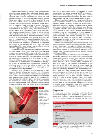
the presence of a foreign body (Figure 4.2). Dental disease occluding the nasopharynx secondary to an inflammatory
can also cause unilateral nasal discharge, and periodontal process such as bacterial or viral rhinotracheitis, trauma
disease should be ruled out during an oral examination. or possibly following nasopharyngeal reflux under general
anaesthesia (Mitten, 1988; De Lorenzi et al., 2015).
4.2
(a) An arrowhead Diagnostics
lodged in the nasal
cavity of a dog The diagnostic evaluation should be guided by clinical
that caused signs observations (Figure 4.3). Evaluation of regional lymph
of chronic nodes is an important part of the physical examination.
mucopurulent
rhinitis is removed The lymph nodes should be evaluated by fine-needle
during a dorsal
rhinotomy. High-yield diagnostics
(b) Close-up of the
arrowhead. • Diagnostic imaging: radiographs, CT, MRI
• Rhinoscopy
• Biopsy
Low-yield diagnostics
(a) • CBC
• Serum chemistry
• Urinalysis
• Cultures: bacterial and fungal
• Coagulation profile
• Fungal serology
• Nasal cytology
Many diagnostics performed whilst working up chronic nasal
4.3
disease do not lead to a specific diagnosis but provide general
(b) health status information. CBC = complete blood count; CT = computed
tomography; MRI = magnetic resonance imaging.
47
Ch04 HNT.indd 47 31/08/2018 10:49