
Page 341 - Avian Virology: Current Research and Future Trends
P. 341
332 | Coppo et al.
Clinical signs lesions may vary from mild mucoid exudation and thickening of
Differing severities of clinical signs and gross lesions has been the mucosa to severe haemorrhages or diphtheritic lesions. Espe-
attributed to two forms of the disease, enzootic and epizootic. cially in mild cases, lesions are more readily detectable in larynx
However, the severity of clinical signs and gross lesions, and the and upper trachea than in other regions of the trachea. Similar
level of morbidity and mortality of ILT, are primarily dependent lesions have also been reported in oral cavity extending to upper
on the immune status of the host, viral strains involved, and pos- oesophagus (Seifried, 1931; Sary et al., 2017). Recent investiga-
sibly the influence of contributing factors such as stress. Recent tions have shown that both the pathogenicity of the infecting
studies have also shown that early exposure to MDV can influ- virus, and the route of viral entry, may influence the anatomic
ence protective immunity against ILTV (Faiz et al., 2016). site of gross and microscopic lesions in immunologically mature
Severe forms of the disease are often seen in susceptible chickens (Beltrán et al., 2017).
(unvaccinated) birds infected with highly virulent virus strains
and are associated with high morbidity (up to 100%) and mortal- Microscopic lesions
ity (approximately 20%) (Noormohammadi and Devlin, 2014). Microscopic lesions may vary depending on the severity and stage
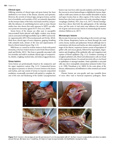
Birds show clinical signs of severe dyspnoea, gasping, breathing of the disease. Respiratory lesions are found through the nasal
with extended neck, closure of the eyes and expectoration of cavity, larynx, trachea and pulmonary bronchi although due to
blood or blood-stained mucus (Fig. 11.2). convenience only larynx and trachea are often examined. At early
Mild forms are caused by virulent strains in birds with partial stages of the disease, respiratory lesions consist of hyperplasia of
immunity, or by less virulent strains in naïve birds (Noormoham- goblet cells and mucus glands, followed by hyperplasia, degen-
madi and Devlin, 2014). This form is generally associated with eration and sloughing of the epithelial cells, and congestion and
low mortality and mild to moderate clinical signs of conjunctivi- rupture of blood capillaries (Fig. 11.3). Exudates consisting of
tis, rhinitis, coughing, tracheal rales, and reduced egg production. sloughed epithelial cells, blood and inflammatory cells are found
in the respiratory lumen. Occasional syncytial cells are also found
Gross lesions in epithelium or amongst exudates. Some epithelial or syncytial
Gross lesions are predominantly found in the conjunctiva and cells may show eosinophilic intranuclear inclusion bodies (Guy
the upper respiratory system (Fig. 11.2). Conjunctival lesions et al., 1990; Timurkaan et al., 2003). In the acute phase of the
may appear in presence or absence of severe lesions in the larynx disease, inflammatory cell infiltration is often minimal and mixed
and trachea, and may consist of serous to mucoid conjunctival in nature.
exudation, occasionally associated with partial to complete clo- Chronic lesions are non-specific and may resemble those
sure of the eyes and thickening of the eyelids. Laryngotracheal of some other viral or bacterial respiratory pathogens. These
Figure 11.2 Common clinical signs (A and B) and lesions (C and D) associated with ILT. (A) Sero-mucoid conjunctivitis and dried exudates
around the eye. (B) Open mouth breathing. (C) Haemorrhagic laryngotracheitis. (D) Diphtheritic laryngotracheitis.