
Page 342 - Avian Virology: Current Research and Future Trends
P. 342
Infectious Laryngotracheitis Virus | 333
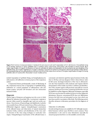
Figure 11.3 Common microscopic lesions in trachea (A-D) and conjunctiva (E and F) associated with ILT. (A) Erosion of the epithelial lining
of the trachea, exposure of blood capillaries and a moderate mainly mononuclear inflammatory cell infiltration in lamina propria. (B) A
higher magnification of image B. (C) Exudation of necrotic epithelial cell debris and heterophils into the tracheal lumen and sloughing of the
epithelial lining. (D) A higher magnification of image C showing a syncytial cell containing eosinophilic intranuclear inclusion bodies (arrow).
(E) Hyperplasia (top) and sloughing (bottom) of the epithelial lining of the conjunctival mucosa. (F) A higher magnification of image E showing
epithelial cells with eosinophilic intranuclear inclusion bodies (arrow).
include hyperplasia of epithelial lining and lymphoplasmacytic convenient and relatively rapid but typical inclusion bodies take
inflammatory cell infiltration in lamina propria of the respiratory time to develop and may disappear before clinical disease takes
system. its full course. Serological detection of the virus in affected tis-
Conjunctival lesions predominantly consist of thickening of sues also bears limitations with sensitivity and requires skill for
the conjunctival mucosa due to hyperplasia of epithelial lining, interpretation of the results (see below). Isolation and identifica-
infiltration of a mixed population of inflammatory cells into tion of the causative agent is still practised, especially for research
lamina propria, syncytial cell formation and rare intranuclear purposes, but the process is time consuming and therefore my not
inclusion bodies. be suitable for routine diagnosis. Molecular techniques, especially
PCR, have now become the method of choice in many diagnostic
Diagnosis laboratories. However conventional PCR does not differentiate
Clinical signs of dyspnoea and gasping may also occur in birds between the field virus and attenuated vaccine virus which may
affected by infectious bronchitis (IB, a coronavirus), respiratory have been used for vaccination of the birds. The following section
mycosis (often caused by Aspergillus spp.) and even ascites syn- describes advances in laboratory procedures for the diagnosis of
drome. Gross lesions of haemorrhagic and diphtheritic tracheitis ILT.
also resemble those of IB and virulent forms of Newcastle disease.
Differential diagnosis requires histopathological examination Detection of antibodies
of the affected tissues and observation of typical intranuclear Several serological assays are available for ILT although enzyme-
inclusion bodies, isolation and identification of the virus and/ linked immuno-sorbent assay (ELISA) is most common. Virus
or detection of the virus using serological or molecular tech- neutralization is also used in some laboratories especially for
niques. Histopathological examination of the affected tissues is research purposes but there is no kit available commercially.