
Page 215 - Canine Lameness
P. 215
13.7 ctrF gesrresres ooracgoe ctrf rFrral reg o 187
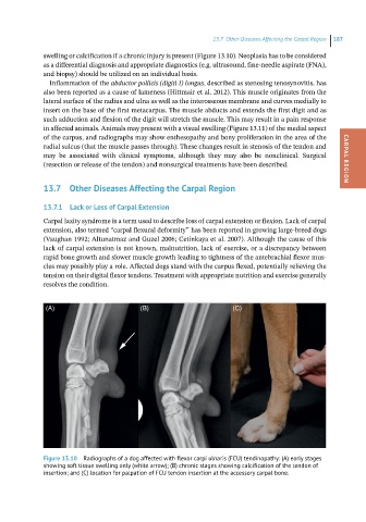
swelling or calcification if a chronic injury is present (Figure 13.10). Neoplasia has to be considered
as a differential diagnosis and appropriate diagnostics (e.g. ultrasound, fine-needle aspirate (FNA),
and biopsy) should be utilized on an individual basis.
Inflammation of the abductor pollicis (digiti I) longus, described as stenosing tenosynovitis, has
also been reported as a cause of lameness (Hittmair et al. 2012). This muscle originates from the
lateral surface of the radius and ulna as well as the interosseous membrane and curves medially to
insert on the base of the first metacarpus. The muscle abducts and extends the first digit and as
such adduction and flexion of the digit will stretch the muscle. This may result in a pain response
in affected animals. Animals may present with a visual swelling (Figure 13.11) of the medial aspect
of the carpus, and radiographs may show enthesopathy and bony proliferation in the area of the
radial sulcus (that the muscle passes through). These changes result in stenosis of the tendon and
may be associated with clinical symptoms, although they may also be nonclinical. Surgical CARPAL REGION
(resection or release of the tendon) and nonsurgical treatments have been described.
13.7 Other Diseases Affecting the Carpal Region
13.7.1 Lack or Loss of Carpal Extension
Carpal laxity syndrome is a term used to describe loss of carpal extension or flexion. Lack of carpal
extension, also termed “carpal flexural deformity” has been reported in growing large-breed dogs
(Vaughan 1992; Altunatmaz and Guzel 2006; Cetinkaya et al. 2007). Although the cause of this
lack of carpal extension is not known, malnutrition, lack of exercise, or a discrepancy between
rapid bone growth and slower muscle growth leading to tightness of the antebrachial flexor mus-
cles may possibly play a role. Affected dogs stand with the carpus flexed, potentially relieving the
tension on their digital flexor tendons. Treatment with appropriate nutrition and exercise generally
resolves the condition.
(A) (B) (C)
Figure 13.10 Radiographs of a dog affected with flexor carpi ulnaris (FCU) tendinopathy: (A) early stages
showing soft tissue swelling only (white arrow); (B) chronic stages showing calcification of the tendon of
insertion; and (C) location for palpation of FCU tendon insertion at the accessory carpal bone.