
Page 154 - Clinical Small Animal Internal Medicine
P. 154
122 Section 3 Cardiovascular Disease
This subjective evaluation provides information on the Evaluation of Fluid Accumulation
VetBooks.ir pulse pressure (i.e. the difference between systemic arte- Right‐sided CHF leads to accumulation of fluid in body
rial peak systolic and diastolic pressures) and potential
cavities, most commonly in the abdomen. The abdomen
presence of various heart diseases.
The quality of pulses in the two limbs should be com- should therefore be palpated for ballottement and the
pared, particularly in cats. Absent femoral arterial pulse in presence of fluid accumulation (ascites). Mild ascites is
difficult to detect, whereas moderate to severe ascites is
one or both limbs may be indicative of arterial thrombo- comparably easy to detect. Pleural effusion is more dif-
embolism. The femoral artery should be palpated simulta- ficult to detect and thoracic radiography or centesis is
neously with cardiac auscultation to detect heartbeats that often required to obtain a definite diagnosis. The limbs
do not produce pulses. Arrhythmias can cause the heart to should also be inspected for the presence of peripheral
contract before it is properly filled with blood, producing edema, although this is an extremely unusual finding in
variable pulses or pulse deficits.
dogs and cats with heart disease.
Palpation of the Precordium
Auscultation
The precordium is evaluated by placing palms and fin-
gers on each side of the thorax over the cardiac ostia Cardiac auscultation is an important component of the
(Figure 15.1). Normally, the cardiac contraction (precor- physical examination in dogs and cats with suspected car-
dial impulse) is best felt on the left side of the thorax over diac disease. An exact diagnosis can seldom be established
the apical area. Changes in cardiac size and shape, or based solely on cardiac auscultation, but optimal ausculta-
space‐occupying thoracic masses can cause a shift of the tion technique can substantially narrow down the list of
location of the precordial impulse. A weak impulse can differential diagnoses. Auscultation allows assessment of
be caused by obesity, hypovolemia, thoracic masses, heart rate, rhythm, and presence of abnormal sounds, such
pleural or pericardial effusion, or pneumothorax. as murmur, click, and additional heart sounds.
Increased precordial impulse can be caused by hyperki- Auscultation should be performed in a quiet environ-
netic conditions such as stress or anemia or conditions ment, and background noise should be minimized. The
characterized by cardiac volume overload and cardio- animal should preferably be standing on all four limbs
megaly, such as severe MMVD or DCM. Severe valvular during cardiac auscultation as abnormal sounds, which
regurgitation or stenosis can cause palpable “buzzing” can easily be mistaken for heart murmurs, can arise in a
vibrations called a precordial thrill. The intensity of the recumbent animal due to rubbing of the heart against the
auscultated heart murmur is always greatest over the chest wall. Furthermore, localization of the area of origin
area of the thrill. of different cardiac sounds is more easily achieved in a
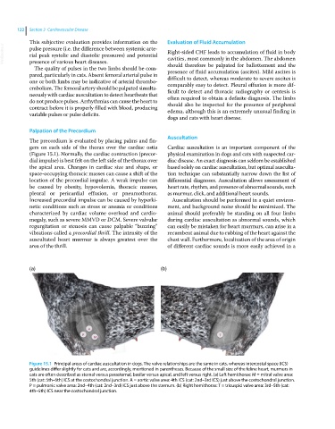
(a) (b)
Figure 15.1 Principal areas of cardiac auscultation in dogs. The valve relationships are the same in cats, whereas intercostal space (ICS)
guidelines differ slightly for cats and are, accordingly, mentioned in parentheses. Because of the small size of the feline heart, murmurs in
cats are often described as sternal versus parasternal, basilar versus apical, and left versus right. (a) Left hemithorax: M = mitral valve area:
5th (cat: 5th–6th) ICS at the costochondral junction. A = aortic valve area: 4th ICS (cat: 2nd–3rd ICS) just above the costochondral junction.
P = pulmonic valve area: 2nd–4th (cat: 2nd–3rd) ICS just above the sternum. (b) Right hemithorax: T = tricuspid valve area: 3rd–5th (cat:
4th–5th) ICS near the costochondral junction.