
Page 155 - Clinical Small Animal Internal Medicine
P. 155
15 Approach to the Patient with Suspected Cardiovascular Disease 123
standing animal. The entire cardiac area should be lesions (such as thoracic masses), diaphragmatic
VetBooks.ir auscultated thoroughly. The areas of auscultation of the hernias, shock, bradycardias, or severely affected myo-
cardial function. The intensity of S2 is commonly
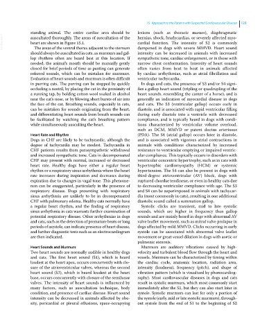
heart are shown in Figure 15.1.
The areas of the ventral thorax adjacent to the sternum
should always be auscultated in cats, as murmurs and gal- dampened in dogs with severe MMVD. Heart sound
intensity can be increased in animals with increased
lop rhythms often are heard best at this location. If sympathetic tone, cardiac enlargement, or in those with
needed, the animal’s mouth should be manually gently narrow chest conformation. Intensity of heart sounds
closed for brief periods of time as panting can generate often varies from beat to beat in animals affected
referred sounds, which can be mistaken for murmurs. by cardiac arrhythmias, such as atrial fibrillation and
Evaluation of heart sounds and murmurs is often difficult ventricular tachycardia.
in purring cats. The purring can be stopped by quickly In dogs and cats, the presence of S3 and/or S4 signi-
occluding a nostril, by placing the cat in the proximity of fies a gallop heart sound (tripling or quadrupling of the
a running tap, by holding cotton wool soaked in alcohol heart sounds, resembling the canter of a horse), and is
near the cat’s nose, or by blowing short bursts of air into generally an indication of myocardial disease in dogs
the face of the cat. Breathing sounds, especially in cats, and cats. The S3 (ventricular gallop) occurs early in
can be mistaken for sounds originating from the heart, diastole, and is associated with rapid ventricular filling
and differentiating heart sounds from breath sounds can during early diastole into a ventricle with decreased
be facilitated by watching the cat’s breathing pattern compliance, and is typically heard in dogs with condi-
while simultaneously ausculting the heart. tions characterized by ventricular volume overload,
such as DCM, MMVD or patent ductus arteriosus
Heart Rate and Rhythm (PDA). The S4 (atrial gallop) occurs later in diastole,
Dogs in CHF are likely to be tachycardic, although the and is associated with vigorous atrial contractions in
degree of tachycardia may be modest. Tachycardia in animals with conditions characterized by increased
CHF patients results from parasympathetic withdrawal resistance to ventricular emptying or impaired ventric-
and increased sympathetic tone. Cats in decompensated ular compliance. This typically occurs in disorders with
CHF may present with normal, increased or decreased ventricular concentric hypertrophy, such as in cats with
heart rate. Healthy dogs have either a regular heart hypertrophic cardiomyopathy (HCM) or systemic
rhythm or a respiratory sinus arrhythmia where the heart hypertension. The S4 can also be present in dogs with
rate increases during inspiration and decreases during third‐degree atrioventricular (AV) block, dogs with
expiration due to changes in vagal tone. This phenome- ruptured chordae tendineae, or even in healthy cats due
non can be exaggerated, particularly in the presence of to decreasing ventricular compliance with age. The S3
respiratory disease. Dogs presenting with respiratory and S4 can be superimposed in animals with tachycar-
sinus arrhythmia are unlikely to be in decompensated dia (most commonly in cats), resulting in one additional
CHF with pulmonary edema. Healthy cats normally have diastolic sound called a summation gallop.
a regular heart rhythm, and the finding of respiratory Systolic clicks are transient, mid to late systolic
sinus arrhythmia in cats warrants further examination of sounds, which are higher in frequency than gallop
potential respiratory disease. Other arrhythmias in dogs sounds and are mainly heard in dogs with abnormal AV
and cats, such as the detection of premature beats or long valve leaflet movement, such as mitral valve prolapse in
periods of asystole, can indicate presence of heart disease, dogs affected by mild MMVD. Clicks occurring in early
and further diagnostic tests such as an electrocardiogram systole can be associated with abnormal valve leaflet
are then indicated. movement or great vessel dilation in dogs with aortic or
pulmonic stenosis.
Heart Sounds and Murmurs Murmurs are auditory vibrations caused by high‐
Two heart sounds are normally audible in healthy dogs velocity and turbulent blood flow through the heart and
and cats. The first heart sound (S1), which is heard vessels. Murmurs can be characterized by timing within
loudest at the heart apex, occurs concurrently with clo- the cardiac cycle, anatomic location, radiation area,
sure of the atrioventricular valves, whereas the second intensity (loudness), frequency (pitch), and shape of
heart sound (S2), which is heard loudest at the heart vibration pattern (which is visualized by phonocardiog-
base, occurs concurrently with closure of the semilunar raphy). Most cardiovascular diseases in dogs and cats
valves. The intensity of heart sounds is influenced by result in systolic murmurs, which most commonly start
many factors, such as auscultation technique, body immediately after the S1, but they can also start later in
condition, and presence of cardiac disease. Heart sound systole. Systolic murmurs can last for only a portion of
intensity can be decreased in animals affected by obe- the systole (early, mid or late systolic murmurs), through-
sity, pericardial or pleural effusions, space‐occupying out systole from the end of S1 to the beginning of S2