
Page 233 - Clinical Small Animal Internal Medicine
P. 233
20 Ventricular Arrhythmias 201
(a)
VetBooks.ir
(b)
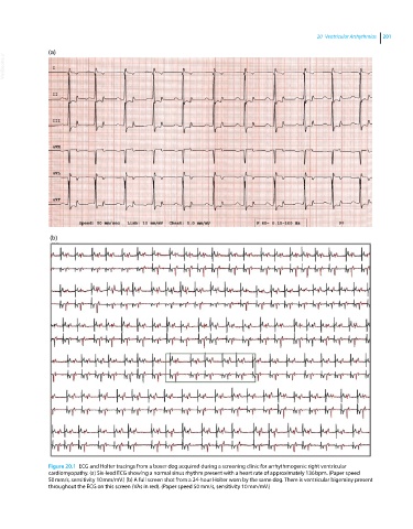
Figure 20.1 ECG and Holter tracings from a boxer dog acquired during a screening clinic for arrhythmogenic right ventricular
cardiomyopathy. (a) Six‐lead ECG showing a normal sinus rhythm present with a heart rate of approximately 136 bpm. (Paper speed
50 mm/s, sensitivity 10 mm/mV.) (b) A full screen shot from a 24‐hour Holter worn by the same dog. There is ventricular bigeminy present
throughout the ECG on this screen (VAs in red). (Paper speed 50 mm/s, sensitivity 10 mm/mV.)