
Page 252 - Clinical Small Animal Internal Medicine
P. 252
220 Section 3 Cardiovascular Disease
Pathophysiology of Target Organ Damage protein‐losing renal disease, hyperadrenocorticism,
VetBooks.ir Arterial blood flow is maintained at relatively constant adrenal tumors, including pheochromocytomas and
aldosterone‐secreting tumors, and diabetes mellitus.
levels in the brain, kidneys, and eyes through a process of
vascular autoregulation, wherein small resistance vessels The most common diseases associated with feline HT
include chronic renal disease, feline hyperthyroidism,
constrict when BP is elevated and dilate when BP is hyperaldosteronism, and diabetes mellitus. In both spe-
decreased to maintain flow. Hypertensive damage in cies, the suspected association (medical or age group
these organs occurs when these autoregulatory mecha- related) between subclinical renal dysfunction and both
nisms fail. Elevated BP without adequate resistance ves- diabetes mellitus and hyperthyroidism may partially
sel constriction causes overdistension of vessels, which account for the occurrence of HT in affected animals.
leads to damage to endothelial tight junctions and allows Ten to 15% of hyperthyroid cats that are normotensive at
protein and plasma leakage into interstitial tissue in the time of diagnosis may become hypertensive after
affected tissue beds (i.e., tissue edema). If excessive vas- successful therapy for hyperthyroidism, indicating other
cular resistance vessel constriction occurs, ischemia of mechanisms of HT at work in these patients. The preva-
local tissues may result in focal hemorrhage and necro- lence of idiopathic and situational HT remains unclear
sis. Permanent structural changes such as arteriosclero- for both species.
sis may develop, decreasing vascular distensibility. Any
combination of these mechanisms may result in clinical
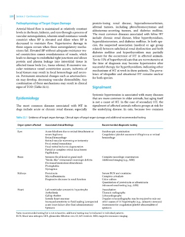
signs of TOD (Table 22.1). Signalment
Systemic hypertension is associated with many diseases
Epidemiology that are more common in older animals, but aging itself
is not a cause of HT. In the case of secondary HT, the
The most common diseases associated with HT in signalment of affected animals reflects groups at risk for
dogs include acute or chronic renal disease, especially the underlying disease. In cats, because two common
Table 22.1 Evidence of target organ damage. Clinical signs of target organ damage and additional recommended testing
Organ system affected Associated clinical findings Recommended diagnostic testing
Eyes Acute blindness due to retinal detachment or Fundoscopic examination
severe hyphema Coagulation/platelet assessment if hyphema or retinal
Retinal hemorrhage hemorrhage
Retinal vascular narrowing or tortuosity
Focal retinal transudates
Focal retinal ischemic degeneration
Partial or complete retinal detachment
Papilledema
Brain Seizures (focal facial or grand mal) Complete neurologic examination
“Stroke‐like” intracranial neurologic deficits Additional imaging (e.g., MRI)
Decreased mentation/obtundation
Photophobia
Nystagmus
Kidneys Proteinuria Serum BUN and creatinine
Microalbuminuria Complete urinalysis
Progressive decrease in renal function Urine culture
Quantitation of proteinuria or albuminuria
Advanced renal testing (e.g., GFR)
Heart Left ventricular concentric hypertrophy Auscultation
Arrhythmia Thoracic radiographs
Gallop rhythm Echocardiography
Systolic heart murmur Doppler echocardiography may be required to rule out
Increased sensitivity to fluid loading (unexpected other causes of LV hypertrophy (e.g., subaortic stenosis)
acute heart failure after fluid administration) Assessment for coagulation/platelet abnormalities if
Epistaxis epistaxis
Note: recommended testing list is not exhaustive, additional testing may be indicated in individual patients.
BUN, blood urea nitrogen; GFR, glomerular filtration rate; LV, left ventricle; MRI, magnetic resonance imaging.