
Page 247 - Clinical Small Animal Internal Medicine
P. 247
21 Supraventricular Arrhythmias 215
ventricular response rate usually approaches the heart flutter results from an electrical impulse circling rapidly
VetBooks.ir rate recorded in cats with sinus rhythm and heart failure. and continuously around a large area of myocardium.
Focal junctional tachycardia is caused by the rapid dis
charge of cells in the area of the AV node. Finally, atrio
Other Forms of Supraventricular ventricular reciprocating tachycardias result from the
Tachyarrhythmia presence of an accessory pathway, which is a small strand
of muscle tissue bridging the atria and ventricles through
Etiology/Pathophysiology the cardiac skeleton and forming an alternative route of
atrioventricular or ventriculoatrial electrical conduction
Initiation of tachyarrhythmias requires a suitable sub besides the atrioventricular node. When conduction is
strate and precipitating factors. Interstitial fibrosis, also possible from atrium to ventricle, preexcitation may
inflammation, ischemia or atrial chamber dilation, com be identified during periods of sinus rhythm as short PR
bined with adrenergic stimulation and electrolyte abnor intervals and a widening of the initial portion of the QRS
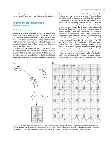
malities promote the risk for tachyarrhythmia. It is complexes (Figure 21.8). This finding confirms the exist
therefore expected that SVTs are commonly diagnosed ence of an accessory pathway. During episodes of atrio
in pets with heart failure. ventricular reciprocating tachycardia, the impulse typically
Supraventricular tachyarrhythmias encompass focal descends along the atrioventricular node to the ventricles
atrial tachycardia, atrial flutter, focal junctional tachycar and returns to the atrium using the accessory pathway.
dia, and atrioventricular reciprocating tachycardia. Focal Sustained tachyarrhythmias can lead to the develop
atrial tachycardia is caused by the rapid and repeated acti ment of heart failure, a phenomenon known as tachycar
vation of a small area of diseased atrial myocytes. Atrial diomyopathy. It is likely that a sustained rate above
(a) (b)
I E 6.0~ 0.5–4.0 H:W
SA node AP
II
AV node
III
aVR
aVL
aVF
Figure 21.8 Ventricular preexcitation. (a) When a dog has an accessory pathway (AP), the sinus impulse may conduct simultaneously
through the atrioventricular node and the abnormal muscle bundle. Ventricular activation is initiated without delay from the accessory
pathway, resulting in a short PR interval and widening of the QRS complex. (b) Six‐lead ECG of a dog with ventricular preexcitation.