
Page 242 - Clinical Small Animal Internal Medicine
P. 242
210 Section 3 Cardiovascular Disease
Diagnosis
VetBooks.ir Electrocardiography
The ECG of persistent atrial standstill is characterized
by a lack of P‐waves with a regular ventricular or AV
nodal escape rhythm, at a rate of 20–60 bpm in dogs. In
cases of transient atrial standstill from hyperkalemia, a
narrowing of the T‐wave and an increase in its ampli
tude occur when plasma potassium concentration
increases above 5.5–6 mmol/L. As potassium concen
tration continues to rise, it leads to bradycardia associ
ated with reduced P‐wave amplitude and a widening
of the QRS complexes. The P‐waves then disappear,
mimicking the ECG of persistent atrial standstill
(Figure 21.5). Another term used to describe this
rhythm disturbance associated with hyperkalemia is
sinoventricular rhythm.
Echocardiography
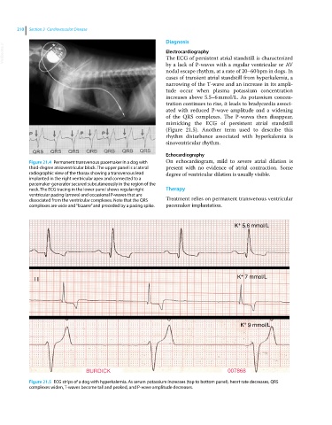
Figure 21.4 Permanent transvenous pacemaker in a dog with On echocardiogram, mild to severe atrial dilation is
third‐degree atrioventricular block. The upper panel is a lateral present with no evidence of atrial contraction. Some
radiographic view of the thorax showing a transvenous lead degree of ventricular dilation is usually visible.
implanted in the right ventricular apex and connected to a
pacemaker generator secured subcutaneously in the region of the
neck. The ECG tracing in the lower panel shows regular right Therapy
ventricular pacing (arrows) and occasional P‐waves that are
dissociated from the ventricular complexes. Note that the QRS Treatment relies on permanent transvenous ventricular
complexes are wide and “bizarre” and preceded by a pacing spike. pacemaker implantation.
+
K 5.6 mmol/L
+
K 7 mmol/L
I I
+
K 9 mmol/L
BURDICK 007868
Figure 21.5 ECG strips of a dog with hyperkalemia. As serum potassium increases (top to bottom panel), heart rate decreases, QRS
complexes widen, T‐waves become tall and peaked, and P‐wave amplitude decreases.