
Page 239 - Clinical Small Animal Internal Medicine
P. 239
21 Supraventricular Arrhythmias 207
VetBooks.ir
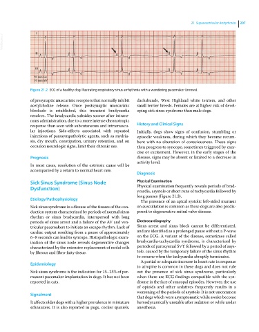
Figure 21.2 ECG of a healthy dog illustrating respiratory sinus arrhythmia with a wandering pacemaker (arrows).
of presynaptic muscarinic receptors that normally inhibit dachshunds, West Highland white terriers, and other
acetylcholine release. Once postsynaptic muscarinic small terrier breeds. Females are at higher risk of devel
blockade is established, this transient bradycardia oping sick sinus syndrome than male dogs.
resolves. The bradycardia subsides sooner after intrave
nous administration, due to a more intense chronotropic
response than seen with subcutaneous and intramuscu History and Clinical Signs
lar injections. Side‐effects associated with repeated Initially, dogs show signs of confusion, stumbling or
injections of parasympatholytic agents, such as mydria episodic weakness, during which they become recum
sis, dry mouth, constipation, urinary retention, and on bent with no alteration of consciousness. These signs
occasion neurologic signs, limit their chronic use. then progress to syncope, sometimes triggered by exer
cise or excitement. However, in the early stages of the
Prognosis disease, signs may be absent or limited to a decrease in
activity level.
In most cases, resolution of the extrinsic cause will be
accompanied by a return to normal heart rate.
Diagnosis
Sick Sinus Syndrome (Sinus Node Physical Examination
Dysfunction) Physical examination frequently reveals periods of brad
ycardia, asystole or short runs of tachycardia followed by
long pauses (Figure 21.3).
Etiology/Pathophysiology The presence of an apical systolic left‐sided murmur
Sick sinus syndrome is a disease of the tissues of the con on auscultation is common as these dogs are also predis
duction system characterized by periods of normal sinus posed to degenerative mitral valve disease.
rhythm or sinus bradycardia, interspersed with long
periods of sinus arrest and a failure of the AV and ven Electrocardiography
tricular pacemakers to initiate an escape rhythm. Lack of Sinus arrest and sinus block cannot be differentiated,
cardiac output resulting from a pause of approximately and are identified as a prolonged pause without a P‐wave
6–8 seconds can lead to syncope. Histopathologic exam on the ECG. A variant of the disease, sometimes called
ination of the sinus node reveals degenerative changes bradycardia‐tachycardia syndrome, is characterized by
characterized by the extensive replacement of nodal cells periods of paroxysmal SVT followed by a period of asys
by fibrous and fibro‐fatty tissue. tole, caused by the temporary failure of the sinus rhythm
to resume when the tachycardia abruptly terminates.
A partial or adequate increase in heart rate in response
Epidemiology
to atropine is common in these dogs and does not rule
Sick sinus syndrome is the indication for 15–25% of per out the presence of sick sinus syndrome, particularly
manent pacemaker implantation in dogs. It has not been when there are ECG findings compatible with the syn
reported in cats. drome in the face of syncopal episodes. However, the use
of opioids and other sedatives frequently results in a
worsening of the periods of asystole. It is not uncommon
Signalment
that dogs which were asymptomatic while awake become
It affects older dogs with a higher prevalence in miniature hemodynamically unstable after sedation or while under
schnauzers. It is also reported in pugs, cocker spaniels, anesthesia.