
Page 240 - Clinical Small Animal Internal Medicine
P. 240
208 Section 3 Cardiovascular Disease
VetBooks.ir
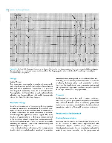
Figure 21.3 Six‐lead ECG of a dog with sick sinus syndrome. After the first two sinus complexes, there is an unexpected 2.3‐second pause
that is terminated by a ventricular escape beat (arrow). Note that the propagation to the ventricles of a P‐wave (*) is prevented by the
occurrence of the escape beat.
Therapy Therefore, atrial pacing when AV nodal function is unaf
fected by disease may be preferred in order to maintain
Medical Therapy synchrony between atrial and ventricular activity.
Two drugs are occasionally successful at temporarily However, the simplest and most commonly used type of
increasing heart rate and alleviating clinical signs in dogs pacing in veterinary patients involves a single lead placed
with sick sinus syndrome. Terbutaline is a selective in the right ventricle via the jugular vein.
beta‐2‐agonist commonly used as a bronchodilator.
Aminophylline or theophylline is a phosphodiesterase
inhibitor and bronchodilator with mild chronotropic Prognosis
effects. See Table 21.2 for drug dosages.
Sudden death is rare in dogs with sick sinus syndrome.
However, long‐term quality of life is typically diminished
Pacemaker Therapy with medical therapy alone. Conversely, permanent
transvenous pacemaker implantation alleviates clinical
Long‐term management of sick sinus syndrome requires signs in the majority of dogs with sick sinus syndrome.
permanent pacemaker implantation. The goal of pace
maker therapy is to stimulate a heartbeat when necessary
in order to maintain the heart rate within a predeter Persistent Atrial Standstill
mined range that optimizes cardiac output. The basic
function of a pacemaker is to deliver an electric current
to the heart that triggers contraction. Modern pacemak Etiology/Pathophysiology
ers also sense the intrinsic cardiac electrical activity, only Persistent atrial standstill, or “silent atrium,” corresponds
delivering an impulse if the native conduction system to the absence of atrial tissue depolarization and
fails to depolarize (Figure 21.4). contraction, despite normal sinus nodal activity.
For optimal cardiac performance, the pacing system Histologically, extensive fibrosis extends throughout the
should mimic normal physiology as closely as possible. atria. It may occur secondary to the progression of