
Page 260 - Clinical Small Animal Internal Medicine
P. 260
228 Section 3 Cardiovascular Disease
VetBooks.ir
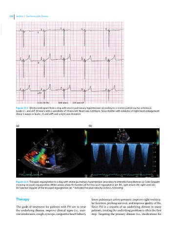
Figure 23.3 Electrocardiogram from a dog with severe pulmonary hypertension secondary to a reverse patent ductus arteriosus.
Leads I, II, and aVF. 50 mm/s with a sensitivity of 10 mm/mV. Heart rate is 85 bpm. Sinus rhythm with evidence of right heart enlargement
(deep S‐waves in leads I, II, and aVF) and a right axis deviation.
(a) (b)
Figure 23.4 Tricuspid regurgitation in a dog with severe pulmonary hypertension secondary to intersitial lung disease. (a) Color Doppler
showing tricuspid regurgitation. White arrows show the borders of the tricuspid regurgitation jet. RA, right atrium; RV, right ventricle.
(b) Spectral Doppler of the tricuspid regurgitation jet. * indicates the peak velocity (4.8 m/s, 92 mmHg).
Therapy lower pulmonary artery pressure, improve right ventricu-
lar function, prolong survival, and improve quality of life.
The goals of treatment for patients with PH are to treat Since PH is a sequela of an underlying disease in many
the underlying disease, improve clinical signs (i.e., exer- patients, treating the underlying problem is often the first
cise intolerance, cough, syncope, congestive heart failure), step. Targeting the primary disease (i.e., medications for