
Page 264 - Clinical Small Animal Internal Medicine
P. 264
232 Section 3 Cardiovascular Disease
(a) (b)
VetBooks.ir
(c) (d)
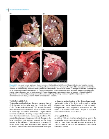
Figure 24.1 Gross postmortem specimens of common congenital heart defects. (a) A dog with patent ductus arteriosus; the image is
taken from a caudal perspective with the descending aorta (Ao) coming toward the viewer. The origin of the ductus (asterisk) within the
aorta can be seen traversing ventral toward the pulmonary artery, which is seen below as the left (L) and right (R) branches. (b) A dog with
tricuspid valve dysplasia showing severe right atrial (RA) enlargement, a sheet‐like tricuspid valve with fused, thick leaflets (arrowheads),
and the ostium of a patent foramen ovale (arrow). (c) A dog with pulmonary valve stenosis showing moderate right ventricular (RV)
hypertrophy and thickened pulmonary valve leaflets (arrow). (d) A dog with a large ventricular septal defect (arrow), seen in the
membranous septum at the junction of the left ventricle (LV) and aorta (Ao).
Ventricular Septal Defects to characterize the location of the defect. Exact confir-
Ventricular septal defects are the most common form of mation of the size of the defect and secondary cardiac
CHD in cats, and are also seen in ~9% of dogs with changes (e.g., aortic insufficiency or left ventricular
CHD. The pathophysiology of VSD is such that small enlargement) carry prognostic information for the
defects (restrictive) may be well tolerated and not result clients. Additionally, some defects may be in a location
in overt signs of cardiac dysfunction. Larger defects, amenable to catheter‐based closure.
however, result in the shunting of large volumes of blood
from the left ventricle to the pulmonary circulation. The Atrial Septal Defects
result of this increased pulmonary flow is damage to the As with a VSD, an atrial septal defect is a hole in the
lung vasculature and volume overload as the blood intracardiac septum separating the left and right heart.
returns to the left heart. VSDs can occur in several dif- ASD is a rare defect in small animals, accounting for
ferent areas along the interventricular septum and 2–6% of cases. In addition, it is often seen in association
numerous classification schemes have been developed with other defects.