
Page 269 - Clinical Small Animal Internal Medicine
P. 269
24 Congenital Heart Disease 237
For dogs, all valve areas should be evaluated, including diastolic in duration, or associated with concurrent
VetBooks.ir the mitral valve area on the left thoracic wall where the api- cardiac signs (arrhythmia, cough, syncope), then CHD
should be considered more likely and further testing
cal impulse is felt most strongly, the semilunar valve area
(aortic and pulmonary valves) at the left heart base (the
ventral 2nd–4th intercostal spaces), the area of the great should be advised.
Other aspects of a cardiac physical examination
vessels just dorsal to the left heart base, and the tricuspid include evaluation of the mucous membranes, jugular
valve area on the right thoracic wall. In the cat, localization veins, and pulse quality. Mucous membranes should be
of individual valve areas is more problematic and ausculta- evaluated for evidence of cyanosis, which may be indica-
tion is usually directed along the left and right sternal bor- tive of a right‐to‐left intracardiac shunt. Very poor
der, moving from caudal to cranial on either side. peripheral perfusion can be suggested by pale mucous
Murmur location and intensity are useful in differenti- membranes and/or a prolonged capillary refill time even
ating between types of CHD as well as for distinguishing in the absence of an obvious heart murmur. Hypokinetic
a physiologic or innocent murmur from that caused by femoral pulses are commonly detected in moderate to
CHD. Left and right apical systolic murmurs are com- severe SAS, while a hyperkinetic pulse quality is sugges-
monly due to atrioventricular valve regurgitation and, in tive of a wide pulse pressure from PDA or severe aortic
a young animal, if an apical systolic murmur is heard, insufficiency. Jugular pulsation with or without disten-
defects of MVD and TVD should be considered most sion can suggest tricuspid regurgitation or elevated
strongly. The murmur of a typical membranous VSD is right‐sided pressures due to tricuspid valve dysplasia,
harsh and systolic at the right sternal boarder. Left basi- PS, or pulmonary hypertension from severe left‐to‐right
lar systolic murmurs can be more problematic as they shunting defects.
can indicate either an innocent murmur or CHD such as
SAS, PS, or ASD. High‐intensity murmurs (grade IV–VI Additional Diagnostic Tests
or louder) usually indicate the presence of significant
CHD regardless of location and are an indication for While the physical examination is an important aspect of
more advanced diagnostics. However, less intense mur- screening for the presence of CHD, it is difficult to make
murs (grade III–VI or less) may originate either from a definitive diagnosis from signalment and physical
CHD or an innocent cause. examination alone. Ancillary diagnostic tests, like tho-
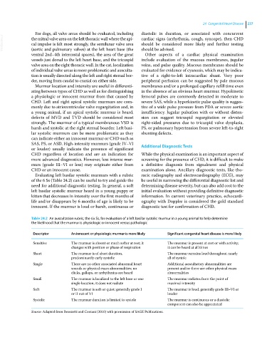
Evaluating left basilar systolic murmurs with a rubric racic radiography and electrocardiography (ECG), may
of the 6 Ss (Table 24.2) can be useful to try and guide the be useful in narrowing the differential diagnostic list and
need for additional diagnostic testing. In general, a soft determining disease severity, but can also add cost to the
left basilar systolic murmur heard in a young puppy or initial evaluation without providing definitive diagnostic
kitten that decreases in intensity over the first months of information. In current veterinary practice, echocardi-
life and/or disappears by 6 months of age is likely to be ography with Doppler is considered the gold standard
innocent. If the murmur is loud or harsh, continuous or diagnostic test for confirmation of CHD.
Table 24.2 An auscultation rubric, the six Ss, for evaluation of a left basilar systolic murmur in a young animal to help determine
the likelihood that the murmur is physiologic or innocent versus pathologic
Descriptor An innocent or physiologic murmur is more likely Significant congenital heart disease is more likely
Sensitive The murmur is absent or much softer at rest; it The murmur is present at rest or with activity;
changes with position or phase of respiration it can be heard at all times
Short The murmur is of short duration, The murmur remains loud throughout nearly
predominantly early systolic all of systole
Single There are no other associated abnormal heart Additional auscultatory abnormalities are
sounds or physical exam abnormalities; no present and/or there are other physical exam
clicks, gallops, or arrhythmias are heard abnormalities
Small The murmur is localized to the left base or one The murmur radiates from the point of
single location, it does not radiate maximal intensity
Soft The murmur is soft or quiet; generally grade I The murmur is loud, generally grade III–VI or
or II out of VI louder
Systolic The murmur duration is limited to systole The murmur is continuous or a diastolic
component can also be appreciated
Source: Adapted from Bronzetti and Corzani (2010) with permission of SAGE Publications.