
Page 273 - Clinical Small Animal Internal Medicine
P. 273
24 Congenital Heart Disease 241
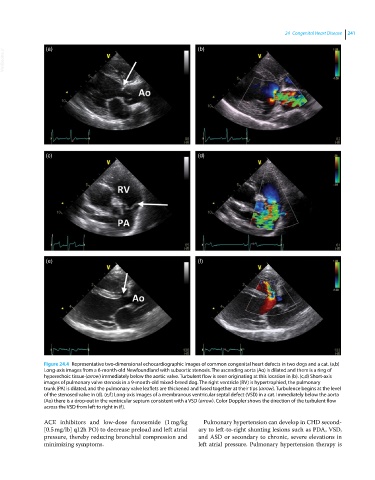
VetBooks.ir (a) (b)
(c) (d)
(e) (f)
Figure 24.4 Representative two‐dimensional echocardiographic images of common congenital heart defects in two dogs and a cat. (a,b)
Long‐axis images from a 6‐month‐old Newfoundland with subaortic stenosis. The ascending aorta (Ao) is dilated and there is a ring of
hyperechoic tissue (arrow) immediately below the aortic valve. Turbulent flow is seen originating at this location in (b). (c,d) Short‐axis
images of pulmonary valve stenosis in a 9‐month‐old mixed‐breed dog. The right ventricle (RV) is hypertrophied, the pulmonary
trunk (PA) is dilated, and the pulmonary valve leaflets are thickened and fused together at their tips (arrow). Turbulence begins at the level
of the stenosed valve in (d). (e,f) Long‐axis images of a membranous ventricular septal defect (VSD) in a cat. Immediately below the aorta
(Ao) there is a drop‐out in the ventricular septum consistent with a VSD (arrow). Color Doppler shows the direction of the turbulent flow
across the VSD from left to right in (f).
ACE inhibitors and low‐dose furosemide (1 mg/kg Pulmonary hypertension can develop in CHD second-
[0.5 mg/lb] q12h PO) to decrease preload and left atrial ary to left‐to‐right shunting lesions such as PDA, VSD,
pressure, thereby reducing bronchial compression and and ASD or secondary to chronic, severe elevations in
minimizing symptoms. left atrial pressure. Pulmonary hypertension therapy is