
Page 271 - Clinical Small Animal Internal Medicine
P. 271
24 Congenital Heart Disease 239
VetBooks.ir (a) (b)
(c) (d)
(e) (f)
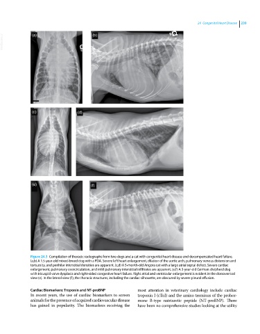
Figure 24.3 Compilation of thoracic radiographs from two dogs and a cat with congenital heart disease and decompensated heart failure.
(a,b) A 1.5‐year‐old mixed‐breed dog with a PDA. Severe left heart enlargement, dilation of the aortic arch, pulmonary venous distension and
tortuosity, and perihilar interstitial densities are apparent. (c,d) A 5‐month‐old Angora cat with a large atrial septal defect. Severe cardiac
enlargement, pulmonary overcirculation, and mild pulmonary interstitial infiltrates are apparent. (e,f) A 3‐year‐old German shepherd dog
with tricuspid valve dysplasia and right‐sided congestive heart failure. Right atrial and ventricular enlargement is evident in the dorsoventral
view (e). In the lateral view (f), the thoracic structures, including the cardiac silhouette, are obscured by severe pleural effusion.
Cardiac Biomarkers: Troponin and NT‐proBNP most attention in veterinary cardiology include cardiac
In recent years, the use of cardiac biomarkers to screen troponin I (cTnI) and the amino terminus of the prohor-
animals for the presence of acquired cardiovascular disease mone B‐type natriuretic peptide (NT‐proBNP). There
has gained in popularity. The biomarkers receiving the have been no comprehensive studies looking at the utility