
Page 270 - Clinical Small Animal Internal Medicine
P. 270
238 Section 3 Cardiovascular Disease
Electrocardiography Thoracic Radiography
VetBooks.ir diac changes related to CHD, though is most useful for plementary information in the evaluation of a patient with
Similar to ECG, thoracic radiographs provide useful sup-
Electrocardiography may be beneficial in detecting car-
suspected CHD. However, thoracic radiography is most
characterizing dysrhythmias heard on auscultation.
CHDs that cause ventricular enlargement, such as PS useful when the veterinarian suspects an animal’s heart
and SAS, may provide a substrate for ventricular disease has decompensated and resulted in pulmonary
arrhythmias secondary to myocardial ischemia. edema or pleural effusion. If signs of respiratory compro-
However, ECG is a more specific rather than sensitive mise or cough are apparent on examination, thoracic radi-
test as animals with CHD may have a normal ECG. ographs are strongly advised. In the asymptomatic animal
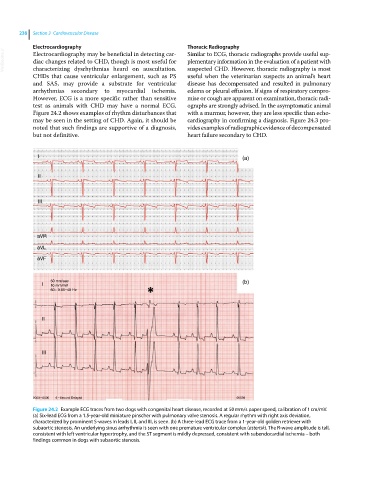
Figure 24.2 shows examples of rhythm disturbances that with a murmur, however, they are less specific than echo-
may be seen in the setting of CHD. Again, it should be cardiography in confirming a diagnosis. Figure 24.3 pro-
noted that such findings are supportive of a diagnosis, vides examples of radiographic evidence of decompensated
but not definitive. heart failure secondary to CHD.
I (a)
II
III
aVR
aVL
aVF
50 mm/sec (b)
I 10 mm/mV
60~ 0.05–40 Hz
II
III
0000–0000 6– Second Delayed 00536
Figure 24.2 Example ECG traces from two dogs with congenital heart disease, recorded at 50 mm/s paper speed, calibration of 1 cm/mV.
(a) Six‐lead ECG from a 1.5‐year‐old miniature pinscher with pulmonary valve stenosis. A regular rhythm with right axis deviation,
characterized by prominent S‐waves in leads I, II, and III, is seen. (b) A three‐lead ECG trace from a 1‐year‐old golden retriever with
subaortic stenosis. An underlying sinus arrhythmia is seen with one premature ventricular complex (asterisk). The R‐wave amplitude is tall,
consistent with left ventricular hypertrophy, and the ST segment is mildly depressed, consistent with subendocardial ischemia – both
findings common in dogs with subaortic stenosis.