
Page 279 - Clinical Small Animal Internal Medicine
P. 279
25 Valvular Heart Disease 247
CHF in 206 dogs with MMVD at different stages. In this regurgitation, a low‐intensity, early systolic murmur
VetBooks.ir study, cough was associated with left atrial enlargement localized on the left apex can be heard. The murmur can
be intermittent and intensity can vary with heart rate
and the presence of an abnormal radiographic airway
pattern. Left atrial enlargement compressing the main-
stem bronchus is believed to induce cough in patients and phase of respiration. When moderate to severe
mitral regurgitation is present, the murmur becomes
with primary bronchomalacia. holosystolic or pansystolic, more intense, and harsher. In
more severe cases, the murmur radiates to the base of
the left thorax and to the right side of the thorax. It
Diagnosis
should be emphasized that the intensity of the murmur
The American College of Veterinary Internal Medicine does not always correlate with the severity of MMVD;
Specialty of Cardiology consensus panel has published that is, loud murmurs do not automatically indicate
guidelines for the diagnosis and treatment of canine severe disease.
chronic valvular heart disease. The guidelines propose a The auscultatory findings can be influenced by several
modification of a staging system for MMVD that is cur- factors such as observer experience, obesity, panting,
rently used to classify human patients with heart failure presence of arrhythmias, and breeds. In a study on pre-
(Table 25.1). This classification introduces the concept of clinical MMVD dogs, 26% of dogs with a confirmed
patients at risk for developing MMVD but that currently echocardiographic diagnosis of MMVD did not have a
do not have heart disease (stage A). The reason for intro- murmur and large‐breed dogs with MMVD have less
ducing this stage is to encourage veterinarians to develop intense murmurs compared with small‐breed dogs.
appropriate screening programs and inform owners Thus, diagnosis of MMVD should not rely only on
regarding the risk of an animal developing the disease. auscultatory findings. Indeed, the ACVIM consensus
Diagnosis of MMVD is generally suspected following a recommends that although the presence of midsystolic
careful auscultation. In early stages of MMVD a midsys- click or left apical murmur in a typical breed is strongly
tolic click may represent the only auscultatory finding. suggestive of MMVD, echocardiographic confirmation
The click is often intermittent and can be better appreci- of the diagnosis is required. Echocardiography can eval-
ated at higher heart rates. It is believed the midsystolic uate the morphology of the valve leaflets and quantify
click is caused by vibration of the prolapsing mitral valve the degree of cardiac enlargement. The echocardio-
leaflets and by the tensing of redundant chordae graphic characteristics of MMVD include prolapse or
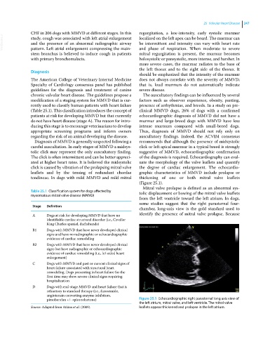
tendineae. In dogs with mild MMVD and mild mitral thickening of one or both mitral valve leaflets
(Figure 25.1).
Mitral valve prolapse is defined as an abnormal sys-
Table 25.1 Classification system for dogs affected by tolic displacement or bowing of the mitral valve leaflets
myxomatous mitral valve disease (MMVD)
from the left ventricle toward the left atrium. In dogs,
some studies suggest that the right parasternal four‐
Stage Definition
chamber, long‐axis view is the gold standard used to
A Dogs at risk for developing MMVD that have no identify the presence of mitral valve prolapse. Because
identifiable cardiac structural disorder (i.e., Cavalier
King Charles spaniel, dachshunds)
B1 Dogs with MMVD that have never developed clinical
signs and have no radiographic or echocardiographic
evidence of cardiac remodeling
B2 Dogs with MMVD that have never developed clinical
signs but have radiographic or echocardiographic
evidence of cardiac remodeling (i.e., lef‐sided heart
enlargement)
C Dogs with MMVD and past or current clinical signs of
heart failure associated with structural heart
remodeling. Dogs presenting in heart failure for the
first time may show severe clinical signs requiring
hospitalization
D Dogs with end‐stage MMVD and heart failure that is
refractory to standard therapy (i.e., furosemide,
angiotensin converting enzyme inhibitors,
pimobendan +/‐ spironolactone) Figure 25.1 Echocardiographic right parasternal long axis view of
the left atrium, mitral valve, and left ventricle. The mitral valve
Source: Adapted from Atkins et al. (2009). leaflets appear thickened and prolapse in the left atrium.