
Page 577 - Clinical Small Animal Internal Medicine
P. 577
50 Diseases of the Oral Cavity and Salivary Glands 545
(a) (b) (c)
VetBooks.ir
(d) (e) (f)
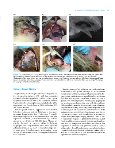
Figure 50.8 Photographs of a 1‐month‐old domestic shorthair with electric burns and extensive tissue necrosis at the lips, cheeks, and
hard palate (a,b). Intraoral dental radiograph of the same patient one year post injury showing the resultant oronasal fistula (c).
Photographs of the same patient one year post injury showing scarring of the palate with a small, right‐sided, persistent oronasal fistula
(arrow) (d); scarring of the cheeks along with developmental enamel and dentin defects of multiple teeth (e); and scarring of the tongue
(f). Copyright 2015 Alexander M. Reiter.
Salivary Gland Disease Sialadenosis typically is a bilateral and painless enlarge
ment of the salivary glands. Although the exact cause of
The prevalence of salivary gland disease in dogs and cats this disease is unknown, concurrent gastrointestinal dis
in a retrospective study was 0.3% , with dogs accounting ease, certain medications, hormonal disorders or primary
for two‐thirds of the population studied. Salivary gland disorders affecting the sympathetic innervation to the
neoplasia accounted for 30% of the cases seen, followed glands have been implicated. Retching and gulping are
by, in order of decreasing frequency, sialadenitis (26%), the most common clinical signs seen with this condition.
degenerative or fibrotic lesions (11%), sialoceles (9%), Differential diagnoses for this disease include inflamma
and infarction (8%). tory variants including necrotizing sialometaplasia, gran
Salivary gland neoplasia appears to have different ulomatous giant cell sialadenosis, and spirocercosis. Both
behaviors in the dog and cat. Cats tend to have more sialadenosis and necrotizing sialometaplasia have shown
advanced disease at the time of diagnosis. A possible response to treatment with phenobarbital at an anticon
familial predisposition in Siamese cats has also been vulsant dose (starting at 2 mg/kg, PO BID). Cases of spi
reported. Despite this, survival times in dogs and cats rocercosis also respond to phenobarbital treatment, but
appear to be similar, at 500–550 days. Masses are this is in addition to macrocyclic lactones used to elimi
usually detected by owners, although halitosis, nate the microorganism. Granulomatous disease has
dysphagia, and exophthalmia have been reported. responded to treatment with glucocorticoids at an anti
Adenocarcinoma is the most common histologic type inflammatory dose. Necrotizing sialometaplasia has been
of tumor seen. A sialolipoma of a minor salivary gland reported in a few cats. In contrast to dogs, excision of the
and lymphosarcoma of major salivary glands have also affected salivary glands in cats provided resolution of
been reported. signs with no recurrence reported.