
Page 573 - Clinical Small Animal Internal Medicine
P. 573
50 Diseases of the Oral Cavity and Salivary Glands 541
with SCC responded to treatment versus 18% of patients collagen degeneration in which degenerated collagen
VetBooks.ir with oral melanoma. The most commonly reported fibers are surrounded by degranulated eosinophils,
resulting in flame figures. Clinically, these lesions are
side‐effect for this combination of treatment was renal
toxicosis, so close monitoring of renal values is
with yellow pinpoint areas consistent with sulfur gran
recommended. characterized by a proliferative or ulcerative component
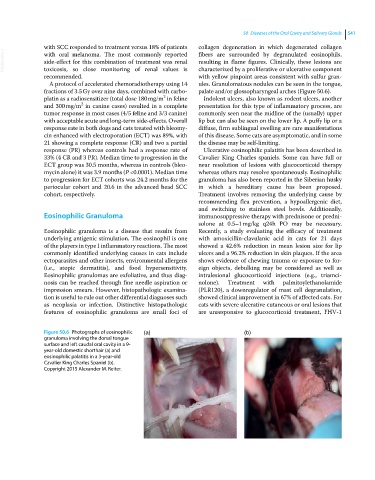
A protocol of accelerated chemoradiotherapy using 14 ules. Granulomatous nodules can be seen in the tongue,
fractions of 3.5 Gy over nine days, combined with carbo palate and/or glossopharyngeal arches (Figure 50.6).
2
platin as a radiosensitizer (total dose 180 mg/m in feline Indolent ulcers, also known as rodent ulcers, another
2
and 300 mg/m in canine cases) resulted in a complete presentation for this type of inflammatory process, are
tumor response in most cases (4/5 feline and 3/3 canine) commonly seen near the midline of the (usually) upper
with acceptable acute and long‐term side‐effects. Overall lip but can also be seen on the lower lip. A puffy lip or a
response rate in both dogs and cats treated with bleomy diffuse, firm sublingual swelling are rare manifestations
cin enhanced with electroporation (ECT) was 89%, with of this disease. Some cats are asymptomatic, and in some
21 showing a complete response (CR) and two a partial the disease may be self‐limiting.
response (PR) whereas controls had a response rate of Ulcerative eosinophilic palatitis has been described in
33% (4 CR and 3 PR). Median time to progression in the Cavalier King Charles spaniels. Some can have full or
ECT group was 30.5 months, whereas in controls (bleo near resolution of lesions with glucocorticoid therapy
mycin alone) it was 3.9 months (P <0.0001). Median time whereas others may resolve spontaneously. Eosinophilic
to progression for ECT cohorts was 24.2 months for the granuloma has also been reported in the Siberian husky
periocular cohort and 20.6 in the advanced head SCC in which a hereditary cause has been proposed.
cohort, respectively. Treatment involves removing the underlying cause by
recommending flea prevention, a hypoallergenic diet,
and switching to stainless steel bowls. Additionally,
Eosinophilic Granuloma immunosuppressive therapy with prednisone or predni
solone at 0.5–1 mg/kg q24h PO may be necessary.
Eosinophilic granuloma is a disease that results from Recently, a study evaluating the efficacy of treatment
underlying antigenic stimulation. The eosinophil is one with amoxicillin‐clavulanic acid in cats for 21 days
of the players in type I inflammatory reactions. The most showed a 42.6% reduction in mean lesion size for lip
commonly identified underlying causes in cats include ulcers and a 96.2% reduction in skin plaques. If the area
ectoparasites and other insects, environmental allergens shows evidence of chewing trauma or exposure to for
(i.e., atopic dermatitis), and food hypersensitivity. eign objects, debulking may be considered as well as
Eosinophilic granulomas are exfoliative, and thus diag intralesional glucocorticoid injections (e.g., triamci
nosis can be reached through fine needle aspiration or nolone). Treatment with palmitoylethanolamide
impression smears. However, histopathologic examina (PLR120), a downregulator of mast cell degranulation,
tion is useful to rule out other differential diagnoses such showed clinical improvement in 67% of affected cats. For
as neoplasia or infection. Distinctive histopathologic cats with severe ulcerative cutaneous or oral lesions that
features of eosinophilic granuloma are small foci of are unresponsive to glucocorticoid treatment, FHV‐1
Figure 50.6 Photographs of eosinophilic (a) (b)
granuloma involving the dorsal tongue
surface and left caudal oral cavity in a 9‐
year‐old domestic shorthair (a) and
eosinophilic palatitis in a 3‐year‐old
Cavalier King Charles Spaniel (b).
Copyright 2015 Alexander M. Reiter.