
Page 572 - Clinical Small Animal Internal Medicine
P. 572
540 Section 6 Gastrointestinal Disease
common oral tumor in dogs. This tumor can vary in different subtypes, including conventional (well differenti
VetBooks.ir appearance and may present as an ulcerated area or a ated, moderately differentiated, and poorly differentiated),
papillary, basaloid, adenosquamous, and spindle cell carci
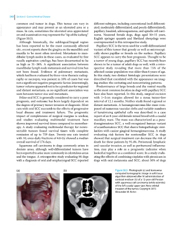
mass. In cats, sometimes the ulcerated area appreciated
on oral examination may represent the “tip of the iceberg”
English springer spaniels and Shetland sheepdogs were
(Figure 50.5). noma. Neutered female dogs, dogs aged 10–15 years,
Although historically, the sublingual/lingual region overrepresented in this retrospective study.
has been reported to be the most commonly affected Papillary SCC is the term used for a well‐differentiated
site, recent reports show the gingiva on the mandible and variant of this tumor that grossly as well as microscopi
maxilla to be most often involved. Metastasis to man cally shows papillae or fronds on the surface. Papillary
dibular lymph nodes in these cases, as evaluated by fine SCC appears to carry the best prognosis. Thought to be
needle aspiration cytology, has been documented to be a tumor of young dogs, papillary SCC has recently been
as high as 31–36%. A significant association between shown to be a tumor of adult dogs as well, with a retro
mandibular lymph node metastasis and survival time has spective study revealing that more than 75% of the
not been found. Evidence of pulmonary metastasis, affected canine population was older than 6 years of age.
which had been evaluated by three‐view thoracic radiog In this study, two distinct histologic presentations were
raphy or necropsy, was present in 10% of cases but was described that correlated with the appearance on imag
not a significant negative prognostic factor. Interestingly, ing studies: the cavitating and noncavitating patterns.
tumor volume appeared not to be a predictor for regional Predominance of large breeds and the rostral maxilla
and distant metastasis, as no significant association was as the most common location in dogs with papillary SCC
seen between tumor size and metastasis. have also been reported. In this study, surgical excision
Feline oral SCC is generally considered to carry a poor with 1–2 cm margins allowed for a mean tumor‐free
prognosis, and outcome has been largely dependent on interval of 12.1 months. Neither study found regional or
the degree of primary tumor invasion at diagnosis. Most distant metastasis. A hemangiosarcoma‐like mass com
cats with oral SCC succumb to the effects of progressive posed of numerous vascular clefts and variable numbers
local disease and treatment failure. The prognostic of keratinizing epithelial cells was described in a case
impact of completeness of surgical margins is unclear, report of an 8‐year‐old female mixed breed with a caudal
and studies evaluating multimodal treatment have maxillary mass. The mass was characterized as a pseu
shown improved survival times compared to monother doangiomatous SCC, a well‐recognized human variant
apy. A study evaluating multimodal therapy for nonre of acanthomatous SCC that shares histopathologic simi
sectable tumors found survival times with complete larities with canine gingival hemangiosarcoma. A study
remission of up to 759 days. Twenty‐one cats treated evaluating risk factors for nontonsillar SCC in dogs
with 10, once‐daily fractions of 4.8‐Gy showed a median showed that surgical treatment can decrease the risk of
overall survival of 174 days. death for these patients by 91.4%. Perineural, lymphatic
Squamous cell carcinoma in dogs commonly arises in and vascular invasion, as well as peritumoral inflamma
dentate areas, although well‐differentiated tumors have tion, may play a role as a prognostic indicator when
been reported to arise more commonly in edentulous areas looked at together as a combined score. In a study evalu
and the tongue. A retrospective study evaluating 84 dogs ating the effects of combining cisplatin with piroxicam in
with a diagnosis of oral and oropharyngeal SCC reported dogs with oral melanoma and SCC, about 50% of dogs
(a) (b) Figure 50.5 Photograph (a) and transverse
computed tomographic image in soft tissue
algorithm obtained after IV administration of
contrast medium (b) of a 12‐year‐old Persian
with squamous cell carcinoma (white asterisks)
of the left caudal upper jaw. Note orbital
invasion of the tumor. Copyright 2015
Alexander M. Reiter.