
Page 861 - Small Animal Clinical Nutrition 5th Edition
P. 861
892 Small Animal Clinical Nutrition
Table 43-1. Common characteristics of canine struvite uroliths.
VetBooks.ir Chemical name Formula Crystal name
Magnesium ammonium
MgNH PO •6H O Struvite
phosphate hexahydrate
4
Variations in mineral composition 4 2
Struvite only
Struvite mixed with lesser quantities of calcium apatite and/or
ammonium acid urate
Nucleus of a different mineral surrounded by variable layers
composed primarily of struvite. Small quantities of calcium
apatite and/or ammonium acid urate also may be present.
Physical characteristics
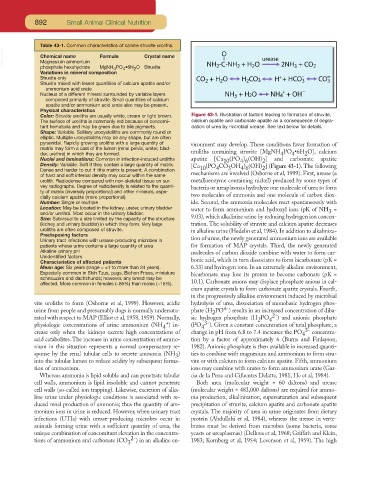
Color: Struvite uroliths are usually white, cream or light brown. Figure 43-1. Illustration of factors leading to formation of struvite,
The surface of uroliths is commonly red because of concomi- calcium apatite and carbonate apatite as a consequence of degra-
tant hematuria and may be green due to bile pigments. dation of urea by microbial urease. See text below for details.
Shape: Variable. Solitary urocystoliths are commonly round or
elliptic. Multiple urocystoliths may be any shape, but are often
pyramidal. Rapidly growing uroliths with a large quantity of vironment may develop. These conditions favor formation of
matrix may form a cast of the lumen (renal pelvis, ureter, blad-
2
4
4
der, urethra) in which they are formed. uroliths containing struvite [MgNH PO •6H O], calcium
Nuclei and laminations: Common in infection-induced uroliths apatite [Ca 10 (PO ) (OH) ] and carbonate apatite
4 6
2
Density: Variable. Soft if they contain a large quantity of matrix. [Ca (PO CO OH ) (OH) ] (Figure 43-1). The following
Dense and harder to cut if little matrix is present. A combination 10 4 3 4 6 2
of hard and soft internal density may occur within the same mechanisms are involved (Osborne et al, 1999). First, urease (a
urolith. Radiodense compared with non-skeletal tissue on sur- metalloenzyme containing nickel) produced by some types of
vey radiographs. Degree of radiodensity is related to the quanti- bacteria or ureaplasmas hydrolyze one molecule of urea to form
ty of matrix (inversely proportional) and other minerals, espe-
cially calcium apatite (more proportional). two molecules of ammonia and one molecule of carbon diox-
Number: Single or multiple ide. Second, the ammonia molecules react spontaneously with
Location: May be located in the kidney, ureter, urinary bladder water to form ammonium and hydroxyl ions (pK of NH =
and/or urethra. Most occur in the urinary bladder. 3
Size: Subvisual to a size limited by the capacity of the structure 9.03), which alkalinize urine by reducing hydrogen ion concen-
(kidney and urinary bladder) in which they form. Very large tration. The solubility of struvite and calcium apatite decreases
uroliths are often composed of struvite. in alkaline urine (Hedelin et al, 1984). In addition to alkaliniza-
Predisposing factors
Urinary tract infections with urease-producing microbes in tion of urine, the newly generated ammonium ions are available
patients whose urine contains a large quantity of urea for formation of MAP crystals. Third, the newly generated
Alkaline urinary pH molecules of carbon dioxide combine with water to form car-
Unidentified factors
Characteristics of affected patients bonic acid, which in turn dissociates to form bicarbonate (pK =
Mean age: Six years (range = ≤1 to more than 24 years). 6.33) and hydrogen ions. In an extremely alkaline environment,
Especially common in Shih Tzus, pugs, Bichon Frises, miniature bicarbonate may lose its proton to become carbonate (pK =
schnauzers and dachshunds; however, any breed may be
affected. More common in females (~85%) than males (~15%). 10.1). Carbonate anions may displace phosphate anions in cal-
cium apatite crystals to form carbonate apatite crystals. Fourth,
in the progressively alkaline environment induced by microbial
vite uroliths to form (Osborne et al, 1999). However, acidic hydrolysis of urea, dissociation of monobasic hydrogen phos-
4-
urine from people and presumably dogs is normally undersatu- phate (H PO ) results in an increased concentration of diba-
2
rated with respect to MAP (Elliot et al, 1958, 1959). Normally, sic hydrogen phosphate (H PO 4 2- ) and anionic phosphate
2
+
physiologic concentrations of urine ammonium (NH ) in- (PO 4 3- ). Given a constant concentration of total phosphate, a
4
crease only when the kidneys excrete high concentrations of change in pH from 6.8 to 7.4 increases the PO 4 3- concentra-
acid catabolites. The increase in urine concentration of ammo- tion by a factor of approximately 6 (Burns and Finlayson,
nium in this situation represents a normal compensatory re- 1982). Anionic phosphate is then available in increased quanti-
sponse by the renal tubular cells to secrete ammonia (NH ) ties to combine with magnesium and ammonium to form stru-
3
into the tubular lumen to reduce acidity by subsequent forma- vite or with calcium to form calcium apatite. Fifth, ammonium
tion of ammonium. ions may combine with urates to form ammonium urate (Gar-
Whereas ammonia is lipid soluble and can penetrate tubular cia de la Pena and Cifuentes Delatte, 1981; He et al, 1984).
cell walls, ammonium is lipid insoluble and cannot penetrate Both urea (molecular weight = 60 daltons) and urease
cell walls (so-called ion trapping). Likewise, excretion of alka- (molecular weight = 483,000 daltons) are required for ammo-
line urine under physiologic conditions is associated with re- nia production, alkalinization, supersaturation and subsequent
duced renal production of ammonia; thus the quantity of am- precipitation of struvite, calcium apatite and carbonate apatite
monium ions in urine is reduced. However, when urinary tract crystals. The majority of urea in urine originates from dietary
infections (UTIs) with urease-producing microbes occur in protein (Abdullahi et al, 1984), whereas the urease in verte-
animals forming urine with a sufficient quantity of urea, the brates must be derived from microbes (some bacteria, some
unique combination of concomitant elevation in the concentra- yeasts or ureaplasmas) (Delluva et al, 1968; Griffith and Klein,
tions of ammonium and carbonate (CO 3 2- ) in an alkaline en- 1983; Kornberg et al, 1954; Levenson et al, 1959). The high