
Page 863 - Small Animal Clinical Nutrition 5th Edition
P. 863
894 Small Animal Clinical Nutrition
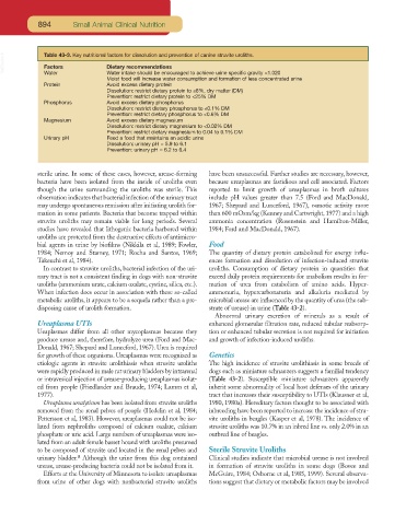
VetBooks.ir Table 43-3. Key nutritional factors for dissolution and prevention of canine struvite uroliths.
Factors
Dietary recommendations
Water
Water intake should be encouraged to achieve urine specific gravity <1.020
Moist food will increase water consumption and formation of less concentrated urine
Protein Avoid excess dietary protein
Dissolution: restrict dietary protein to ≤8%, dry matter (DM)
Prevention: restrict dietary protein to <25% DM
Phosphorus Avoid excess dietary phosphorus
Dissolution: restrict dietary phosphorus to ≤0.1% DM
Prevention: restrict dietary phosphorus to <0.6% DM
Magnesium Avoid excess dietary magnesium
Dissolution: restrict dietary magnesium to <0.02% DM
Prevention: restrict dietary magnesium to 0.04 to 0.1% DM
Urinary pH Feed a food that maintains an acidic urine
Dissolution: urinary pH = 5.9 to 6.1
Prevention: urinary pH = 6.2 to 6.4
sterile urine. In some of these cases, however, urease-forming have been unsuccessful. Further studies are necessary, however,
bacteria have been isolated from the inside of uroliths even because ureaplasmas are fastidious and cell associated. Factors
though the urine surrounding the uroliths was sterile. This reported to limit growth of ureaplasmas in broth cultures
observation indicates that bacterial infection of the urinary tract include pH values greater than 7.5 (Ford and MacDonald,
may undergo spontaneous remission after initiating urolith for- 1967; Shepard and Lunceford, 1967), osmotic activity more
mation in some patients. Bacteria that become trapped within than 600 mOsm/kg (Kenney and Cartwright, 1977) and a high
struvite uroliths may remain viable for long periods. Several ammonia concentration (Rosenstein and Hamilton-Miller,
studies have revealed that lithogenic bacteria harbored within 1984; Ford and MacDonald, 1967).
uroliths are protected from the destructive effects of antimicro-
bial agents in urine by biofilms (Nikkila et al, 1989; Fowler, Food
1984; Nemoy and Stamey, 1971; Rocha and Santos, 1969; The quantity of dietary protein catabolized for energy influ-
Takeuchi et al, 1984). ences formation and dissolution of infection-induced struvite
In contrast to struvite uroliths, bacterial infection of the uri- uroliths. Consumption of dietary protein in quantities that
nary tract is not a consistent finding in dogs with non-struvite exceed daily protein requirements for anabolism results in for-
uroliths (ammonium urate, calcium oxalate, cystine, silica, etc.). mation of urea from catabolism of amino acids. Hyper-
When infection does occur in association with these so-called ammonuria, hypercarbonaturia and alkaluria mediated by
metabolic uroliths, it appears to be a sequela rather than a pre- microbial urease are influenced by the quantity of urea (the sub-
disposing cause of urolith formation. strate of urease) in urine (Table 43-2).
Abnormal urinary excretion of minerals as a result of
Ureaplasma UTIs enhanced glomerular filtration rate, reduced tubular reabsorp-
Ueaplasmas differ from all other mycoplasmas because they tion or enhanced tubular secretion is not required for initiation
produce urease and, therefore, hydrolyze urea (Ford and Mac- and growth of infection-induced uroliths.
Donald, 1967; Shepard and Lunceford, 1967). Urea is required
for growth of these organisms. Ureaplasmas were recognized as Genetics
etiologic agents in struvite urolithiasis when struvite uroliths The high incidence of struvite urolithiasis in some breeds of
were rapidly produced in male rat urinary bladders by intrarenal dogs such as miniature schnauzers suggests a familial tendency
or intravesical injection of urease-producing ureaplasmas isolat- (Table 43-2). Susceptible miniature schnauzers apparently
ed from people (Friedlander and Braude, 1974; Lamm et al, inherit some abnormality of local host defenses of the urinary
1977). tract that increases their susceptibility to UTIs (Klausner et al,
Ureaplasma urealyticum has been isolated from struvite uroliths 1980, 1980a). Hereditary factors thought to be associated with
removed from the renal pelves of people (Hedelin et al, 1984; inbreeding have been reported to increase the incidence of stru-
Pettersson et al, 1983). However, ureaplasmas could not be iso- vite uroliths in beagles (Kasper et al, 1978). The incidence of
lated from nephroliths composed of calcium oxalate, calcium struvite uroliths was 10.7% in an inbred line vs. only 2.0% in an
phosphate or uric acid. Large numbers of ureaplasmas were iso- outbred line of beagles.
lated from an adult female basset hound with uroliths presumed
to be composed of struvite and located in the renal pelves and Sterile Struvite Uroliths
a
urinary bladder. Although the urine from this dog contained Clinical studies indicate that microbial urease is not involved
urease, urease-producing bacteria could not be isolated from it. in formation of struvite uroliths in some dogs (Bovee and
Efforts at the University of Minnesota to isolate ureaplasmas McGuire, 1984; Osborne et al, 1985, 1999). Several observa-
from urine of other dogs with nonbacterial struvite uroliths tions suggest that dietary or metabolic factors may be involved