
Page 833 - Small Animal Clinical Nutrition 5th Edition
P. 833
864 Small Animal Clinical Nutrition
in dogs has not been reported. However, there is a role for
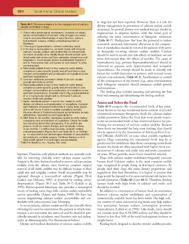
Table 40-7. Recommendations for the management of calcium dietary management in prevention of calcium oxalate urolith
VetBooks.ir oxalate urolithiasis in dogs. recurrence. In general, dietary and medical therapy should be
1. Obtain data (postsurgical radiography, complete urinalysis,
serum concentrations of calcium, urea nitrogen and creati- implemented in stepwise fashion, with the initial goal of
nine) to evaluate effectiveness of renal function, calcium reducing the urine concentration of lithogenic substances
homeostasis, surgery, voiding urohydropropulsion or (Table 40-7). Medications that have the potential to induce
lithotripsy. unwanted, sustained, detrimental alterations in the composi-
2. If the dog is hypercalcemic, correct underlying cause.
3. If the dog is normocalcemic, consider foods with reduced tion of metabolites should be reserved for patients with active
calcium, oxalate, sodium and protein that do not promote or frequently recurring calcium oxalate uroliths. Caution
formation of acidic urine. Ideally foods should contain addi- should be used to ensure that side effects of treatment are not
tional water and citrate and have adequate phosphorus and
magnesium. Avoid excess and/or supplemental vitamins C more detrimental than the effects of uroliths. The cause of
and D. Prescription Diet u/d Canine or w/d Canine* is often hypercalcemia (e.g., primary hyperparathyroidism) should be
recommended. corrected in patients with hypercalcemia and resorptive
4. Reevaluate patient in two to four weeks to verify dietary
compliance (urine specific gravity and pH and serum urea hypercalciuria. An attempt should be made to identify risk
nitrogen concentration) and amelioration of crystalluria (urine factors for urolith formation in patients with normal serum
sediment examination). calcium concentrations (Table 40-2). Amelioration or control
5. Consider additional potassium citrate if calcium oxalate
crystals and aciduria persist. of the consequences of risk factors (e.g., urine oversaturation
6. Reevaluate patient in two to four weeks to verify dietary with lithogenic minerals) should minimize urolith growth
compliance (urine specific gravity and pH and serum urea and recurrence.
nitrogen concentration) and amelioration of crystalluria (urine
sediment examination). Consider vitamin B supplementation The feeding plan includes assessing and selecting the best
6
(2 to 4 mg/kg body weight q24 to 48 hours) if calcium food and assessing and determining the feeding method.
oxalate crystalluria persists.
7. Again, reevaluate patient in two to four weeks to verify Assess and Select the Food
dietary compliance and amelioration of crystalluria. Consider
administration of hydrochlorothiazide (2 mg/kg body weight Table 40-8 compares the recommended levels of key nutri-
q24 to 48 hours) if calcium oxalate crystalluria persists. tional factors to the key nutritional factor content of selected
Adverse effects of hydrochlorothiazide administration include
dehydration, hypokalemia and hypercalcemia. commercial veterinary therapeutic foods for calcium oxalate
8. After three to six months, reevaluate patient to verify dietary urolith prevention. Select the food that most closely match-
compliance and amelioration of crystalluria. Check for urolith es the recommended levels of key nutritional factors for pre-
recurrence by abdominal radiography. If no uroliths are
present, continue current therapy and reevaluate in three venting the recurrence of calcium oxalate uroliths. Because
to six months. If uroliths have recurred, consider voiding these foods are intended for long-term feeding, they should
urohydropropulsion (Figure 38-5 and Table 38-7), or lithotrip- also be approved by the Association of American Feed Con-
sy. If unsuccessful and clinical signs referable to urocystoliths
are persistent, consider surgery. Continue therapy to mini- trol Officials (AAFCO), or some other credible regulatory
mize urolith growth if clinical signs are not present. agency. Dogs consuming dry commercial foods may be at
*Hill’s Pet Nutrition, Inc., Topeka, KS, USA. greater risk for urolithiasis than those consuming moist foods
because dry foods are often associated with higher urine con-
centrations of calcium and oxalic acid and more concentrat-
reported. Therefore, only physical methods are currently avail- ed urine. When possible, moist foods should be selected.
able for removing clinically active calcium oxalate uroliths. Dogs with calcium oxalate urolithiasis frequently consume
Surgery is the time-honored method to remove calcium oxalate human food. Calcium oxalate is the most common urolith
uroliths from the urinary tract; however, complete surgical type recognized in people living in developed countries. As
removal of all visible uroliths may be difficult because of their people feed their dogs the same dietary proportions and
small size and irregular contour. Small urocystoliths may be ingredients they feed themselves, it is logical to assume that
aspirated through a transurethral catheter (Figure 38-6) dogs would be exposed to the same nutritional risk factors for
(Lulich and Osborne, 1992) or removed by voiding urohy- urolith formation (Tables 40-2 and 40-3). Therefore, feeding
dropropulsion (Figure 38-5 and Table 38-7) (Lulich et al, human foods with high levels of calcium and oxalic acid
1993). Extracorporeal lithotripsy also provides a nonsurgical should be avoided.
means of treating some dogs with calcium oxalate nephroliths In addition to consumption of human food, an association
and/or ureteroliths (Adams and Senior, 1999). We have had between calcium oxalate urolithiasis and consumption of
success fragmenting calcium oxalate urocystoliths and ure- commercially available treats has been noted. The high sodi-
throliths with intracorporeal laser lithotripsy. um content of some commercial dog treats may help explain
In some patients, calcium oxalate uroliths are clinically silent, this association because sodium consumption promotes
obviating the need for intervention. For patients in which inter- hypercalciuria (Lulich et al, 1992). Like foods, treats should
vention is not warranted, the status of uroliths should be peri- not contain more than 0.3% DM sodium and they should be
odically assessed by urinalyses, renal function tests and radiog- limited to less than 10% of the total food regimen (volume or
raphy or ultrasonography. (See Reassessment below.) weight basis).
Dietary and medical dissolution of calcium oxalate uroliths Feeding foods designed to dissolve struvite uroliths provides