
Page 828 - Small Animal Clinical Nutrition 5th Edition
P. 828
Canine Calcium Oxalate Urolithiasis 859
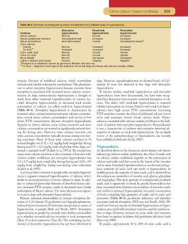
VetBooks.ir Table 40-4. Summary of distinguishing clinical manifestations for different types of hypercalciuria.
Resorptive
Absorptive
Renal-leak
Features
Increased
Serum calcium hypercalciuria hypercalciuria hypercalciuria
Normal
Normal
Serum parathyroid hormone Decreased/normal Increased Increased
Serum phosphorus Normal/increased Normal Decreased/increased*
Urine calcium
Fasting Normal Increased Increased
Dx food** Increased Increased Increased
Urine oxalic acid Normal Normal Normal
Urine uric acid Normal Normal Normal
Bone density Normal Decreased Decreased
Calcium balance (total body) Positive Negative Negative
*Phosphorus is retained in serum as glomerular filtration rate declines.
**Dx food = diagnostic food used in the evaluation of normal dogs and those with calcium oxalate uroliths.
merular filtration of mobilized calcium, which overwhelms dogs. However, hypophosphatemia or elevated levels of 1,25-
normal renal tubular reabsorptive mechanisms.This phenome- vitamin D were not observed in five dogs with absorptive
non is called resorptive hypercalciuria because excessive bone hypercalciuria.
resorption is associated with increased serum calcium concen- In human studies, renal-leak hypercalciuria and resorptive
trations. In dogs, normocalcemic hypercalciuria is thought to hypercalciuria have been documented, but have been recog-
result from either intestinal hyperabsorption of calcium (so- nized less frequently than excessive intestinal absorption of cal-
called absorptive hypercalciuria), or decreased renal tubular cium. The defect with renal-leak hypercalciuria is impaired
reabsorption of calcium (so-called renal-leak hypercalciuria) tubular reabsorption of calcium. Patients with renal-leak hyper-
(Table 40-4). Absorptive hypercalciuria is characterized by calciuria have high serum PTH concentrations. Increasing
increased urine calcium excretion and urine calcium concentra- PTH secretion counters the effect of additional calcium lost in
tion, normal serum calcium concentration and normal or low urine and maintains normal blood calcium levels. Hyper-
serum PTH concentration. Because absorptive hypercalciuria calcemia associated with calcium oxalate urolithiasis is the hall-
depends on dietary calcium, urine calcium excretion and urine mark of patients with resorptive hypercalciuria. Hypercalcemia
calcium concentration are normal or significantly reduced dur- is not a characteristic of patients with excessive intestinal ab-
ing the fasting state. However, urine calcium excretion and sorption of calcium or renal-leak hypercalciuria. An in-depth
urine calcium concentration typically increase during non-fast- review of the pathophysiology of hypercalciuria has recently
ing conditions. Mean 24-hour urine calcium excretion in 33 been published (Park and Pearle, 2007).
normal beagles was 0.32 ± 0.2 mg/kg body weight/day during
fasting and 0.51 ± 0.3 mg/kg body weight/day when dogs con- Hyperoxaluria
a
sumed a standard food (Lulich et al, 1991a). By comparison, As described above in the discussion about dietary risk factors
mean urine calcium excretion in five miniature schnauzers with influencing calcium oxalate urolithiasis, the effect of oxalic acid
calcium oxalate urolithiasis and absorptive hypercalciuria was on calcium oxalate urolithiasis depends on the interactions of
1.0 ± 0.5 mg/kg body weight/day during fasting and 2.84 ± 0.9 calcium and oxalic acid that occur in the lumen of the intestine
mg/kg body weight/day during non-fasting urine collections and in urine. Intestinal hyperabsorption or accelerated endoge-
(Lulich et al, 1991). nous synthesis of oxalic acid can result in hyperoxaluria. In
A primary defect observed in people with absorptive hypercal- healthy people, the majority of urine oxalic acid is derived from
ciuria is apparent intestinal hyperabsorption of calcium, which the endogenous metabolism of ascorbic acid, glycine, glyoxylate
results in increased excretion of excess calcium in urine. In addi- and tryptophan. The daily quantity of endogenously produced
tion to enhanced glomerular filtration of absorbed dietary calci- oxalic acid is apparently minimal. In people, hyperoxaluria has
um, decreased PTH secretion results in decreased renal tubular been associated with inherited abnormalities of excessive oxalic
reabsorption of filtered calcium.The same phenomenon appears acid synthesis (primary hyperoxaluria), increased consumption
to occur in dogs with absorptive hypercalciuria. of foods containing high quantities of oxalic acid or oxalic acid
Primary intestinal abnormalities of calcium absorption, dis- precursors (Table 40-3), pyridoxine deficiency and disorders
orders of 1,25-vitamin D production and hypophosphatemia- associated with fat absorption (Williams and Smith, 1983).We
induced hypervitaminosis D have been recognized as causes of could not find any reports of inherited hyperoxaluria or hyper-
hypercalciuria in people (Park and Pearle, 2007). Absorptive oxaluria associated with intestinal resection and fat malabsorp-
hypercalciuria in people has recently been further subclassified tion in dogs. However, increases in urine oxalic acid excretion
as to whether increased calcium excretion is food unresponsive have been recognized in kittens fed pyridoxine-deficient foods
(Type 1) or food responsive (Type II). The underlying mecha- (Bai et al, 1989).
nism(s) of absorptive hypercalciuria has not been identified in In people, approximately 10 to 20% of urine oxalic acid is