
Page 826 - Small Animal Clinical Nutrition 5th Edition
P. 826
Canine Calcium Oxalate Urolithiasis 857
urolith formation (Tables 40-2 and 40-3).Therefore,reduction
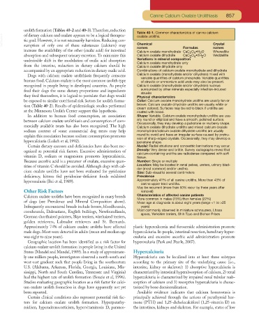
Table 40-1. Common characteristics of canine calcium
VetBooks.ir tic goal. However, it is not necessarily harmless. Reducing con- Chemical Crystal
of dietary calcium and oxalate appears to be a logical therapeu-
oxalate uroliths.
sumption of only one of these substances (calcium) may
names Formulas names
increase the availability of the other (oxalic acid) for intestinal Calcium oxalate monohydrate CaC O •H O Whewellite
2
2 4
absorption and subsequent urinary excretion. To minimize this Calcium oxalate dihydrate CaC O •2H O Weddellite
2 4
2
undesirable shift in the modulation of oxalic acid absorption Variations in mineral composition
Calcium oxalate monohydrate only
from the intestine, reduction in dietary calcium should be Calcium oxalate dihydrate only
accompanied by an appropriate reduction in dietary oxalic acid. Combinations of calcium oxalate monohydrate and dihydrate
Dogs with calcium oxalate urolithiasis frequently consume Calcium oxalate (monohydrate and/or dihydrate) mixed with
variable quantities of calcium phosphate. Variable quantities
human food. Calcium oxalate is the most common urolith type of struvite or ammonium acid urate may also be present.
recognized in people living in developed countries. As people Calcium oxalate (monohydrate and/or dihydrate) nucleus
feed their dogs the same dietary proportions and ingredients surrounded by other minerals especially infection-induced
struvite
they feed themselves, it is logical to postulate that dogs would Physical characteristics
be exposed to similar nutritional risk factors for urolith forma- Color: Calcium oxalate monohydrate uroliths are usually tan or
tion (Table 40-2). Results of epidemiologic studies performed brown. Calcium oxalate dihydrate uroliths are usually white or
cream colored. Surfaces may be red to black if uroliths are
at the Minnesota Urolith Center support this hypothesis. coated with blood.
In addition to human food consumption, an association Shape: Variable. Calcium oxalate monohydrate uroliths are usu-
between calcium oxalate urolithiasis and consumption of com- ally round or elliptical and have a smooth, polished surface.
Occasionally, they may develop a jackstone or mulberry shape.
mercially available treats has also been recognized. The high Calcium oxalate dihydrate uroliths and mixed calcium oxalate
sodium content of some commercial dog treats may help monohydrate/calcium oxalate dihydrate uroliths are usually
explain this association because sodium consumption promotes round to ovoid and have an irregular surface caused by protru-
sion of sharp-edged crystals. Occasionally, they may develop a
hypercalciuria (Lulich et al, 1992). jackstone shape.
Certain dietary excesses and deficiencies have also been rec- Nuclei: Radial striations and concentric laminations may occur.
ognized as potential risk factors. Excessive administration of Density: Very dense and brittle. Survey radiographs reveal that
calcium-containing uroliths are radiodense compared with soft
vitamin D, sodium or magnesium promotes hypercalciuria. tissue.
Because ascorbic acid is a precursor of oxalate, excessive quan- Number: Single or multiple
tities of vitamin C should be avoided. Although dogs with cal- Location: May be located in renal pelves, ureters, urinary blad-
der (most common) and/or urethra.
cium oxalate uroliths have not been evaluated for pyridoxine Size: Sub-visual to several centimeters
deficiency, kittens fed pyridoxine-deficient foods exhibited Prevalence
hyperoxaluria (Bai et al, 1989). Approximately 41% of all canine uroliths. More than 43% of
canine upper tract uroliths.
May be recurrent (more than 50% recur by three years after
Other Risk Factors removal)
Calcium oxalate uroliths have been recognized in many breeds Characteristics of affected canine patients
More common in males (73%) than females (22%)
of dogs (see Prevalence and Mineral Composition above). Mean age at diagnosis is about eight years (range <1 to >25
Infrequently encountered breeds include boxers, bloodhounds, years)
coonhounds, Dalmatians, English bulldogs, Newfoundlands, Most commonly observed in miniature schnauzers, Lhasa
apsos, Yorkshire terriers, Shih Tzus and Bichon Frises
German shorthaired pointers, Skye terriers, wirehaired terriers,
golden retrievers, Labrador retrievers and St. Bernards.
Approximately 74% of calcium oxalate uroliths have affected plastic hypercalcemia and furosemide administration promote
male dogs. Most were detected in adults (mean and median age hypercalciuria. In people, intestinal resection, hereditary hyper-
was eight to nine years). oxaluria and excessive ascorbic acid administration promote
Geographic location has been identified as a risk factor for hyperoxaluria (Park and Pearle, 2007).
calcium oxalate urolith formation in people living in the United
States (Mandel and Mandel, 1989). In a study of approximate- Hypercalciuria
ly one million people, investigators observed a north-south and Hypercalciuria can be localized into at least three subtypes
west-east gradient such that people living in the southeastern according to the primary site of the underlying cause (i.e.,
U.S. (Alabama, Arkansas, Florida, Georgia, Louisiana, Mis- intestine, kidney or skeleton): 1) absorptive hypercalciuria is
sissippi, North and South Carolina, Tennessee and Virginia) characterized by intestinal hyperabsorption of calcium, 2) renal
had the highest rate of urolith formation (Soucie et al, 1996). hypercalciuria is characterized by impaired renal tubular reab-
Studies evaluating geographic location as a risk factor for calci- sorption of calcium and 3) resorptive hypercalciuria is charac-
um oxalate urolith formation in dogs have apparently not yet terized by bone demineralization.
been reported. Available evidence indicates that calcium homeostasis is
Certain clinical conditions also represent potential risk fac- principally achieved through the actions of parathyroid hor-
tors for calcium oxalate urolith formation. Hyperparathy- mone (PTH) and 1,25-dicholecalciferol (1,25-vitamin D) on
roidism, hyperadrenocorticism, hypervitaminosis D, paraneo- the intestines, kidneys and skeleton. For example, states of low