
Page 825 - Small Animal Clinical Nutrition 5th Edition
P. 825
856 Small Animal Clinical Nutrition
VetBooks.ir
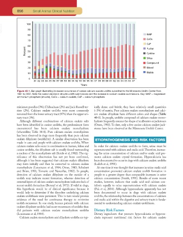
Figure 40-1. Bar graph illustrating increased occurrence of canine calcium oxalate uroliths submitted to the Minnesota Urolith Center from
1981 to 2007. Note the overall decline in struvite urolith submissions and the increase in calcium oxalate submissions. Key: MAP = magnesium
ammonium phosphate (struvite), CaOx = calcium oxalate, CaP = calcium phosphate.
miniature poodles (3%), Chihuahuas (2%) and Jack Russell ter- ically dense and brittle; they have relatively small quantities
riers (2%). Calcium oxalate uroliths were more commonly (~3%) of matrix. Pure calcium oxalate monohydrate and calci-
removed from the lower urinary tract (97%) than the upper uri- um oxalate dihydrate have different colors and shapes (Table
nary tract (3%). 40-1). In people, uroliths composed of calcium oxalate mono-
Although different combinations of calcium oxalate salts hydrate frequently assume the shape of mulberries or jackstones
have been identified in canine uroliths, the predominant form (Otnes, 1983). To date, only a few canine calcium oxalate jack-
encountered has been calcium oxalate monohydrate stones have been observed at the Minnesota Urolith Center.
(whewellite; Table 38-8). Pure calcium oxalate monohydrate
has been observed in dogs more frequently than pure calcium
oxalate dihydrate (weddelite). A similar observation has been ETIOPATHOGENESIS AND RISK FACTORS
made in cats and people with calcium oxalate uroliths. When
calcium oxalate salts occur in combination in human, feline and In order for calcium oxalate uroliths to form, urine must be
canine uroliths, the dihydrate salt is usually found surrounding supersaturated with calcium and oxalic acid.Therefore, increas-
a nucleus of the monohydrate salt (Koide et al, 1982). The sig- ing the urine concentration of calcium and/or oxalic acid pro-
nificance of this observation has not yet been confirmed, motes calcium oxalate crystal formation. Hypercalciuria has
although it has been suggested that calcium oxalate dihydrate been documented to occur in dogs with calcium oxalate uroliths
may form initially and then be converted to calcium oxalate (Lulich et al, 1991).
monohydrate (Leusmann et al, 1984; Otnes, 1983; Schubert At one time it was thought that increases in urine oxalic acid
and Brien, 1981; Tomazic and Nancollas, 1982). In people, concentration promoted calcium oxalate urolith formation in
detection of calcium oxalate dihydrate on the outside of a people to a greater degree than comparable increases in urine
urolith may indicate recent formation, whereas detection of calcium concentration (Smith, 1991). Results of more recent
external layers of calcium oxalate monohydrate indicates lack of studies, however, indicate that oxalic acid and calcium con-
recent urolith formation (Berenyl et al, 1972). If valid in dogs, tribute equally to urine supersaturation with calcium oxalate
this hypothesis would be of clinical significance because it (Pak et al, 2004). Although hyperoxaluria apparently has not
would help to determine if the disorders underlying calcium been documented to occur in dogs with calcium oxalate
oxalate urolithiasis were persistent. This in turn would provide uroliths, the relationship between the concentrations of calcium
evidence of the need for continuous therapy to minimize and oxalic acid within the digestive and urinary tracts is funda-
urolith recurrence. In one study, human patients with calcium mental to understanding calcium oxalate urolithiasis.
oxalate dihydrate uroliths had more recurrences of uroliths than
did patients with calcium oxalate monohydrate uroliths Dietary Risk Factors
(Leusmann et al, 1984). Dietary ingredients that promote hypercalciuria or hyperox-
Calcium oxalate monohydrate and dihydrate uroliths are typ- aluria represent nutritional risk factors for calcium oxalate