
Page 821 - Small Animal Clinical Nutrition 5th Edition
P. 821
Canine Purine Urolithiasis 851
VetBooks.ir Further Discussion
Ammonium urate uroliths are highly recurrent, so prophylactic
therapy should always be considered. Use of a food that avoids
excessive levels of dietary purines and promotes formation of
dilute, alkaline urine is effective in preventing recurrence of
ammonium urate uroliths approximately 80% of the time.
Allopurinol has been recommended for preventive therapy; how-
ever, recent studies indicate that prolonged administration of
high doses of allopurinol may result in formation of xanthine
uroliths. The risk of xanthine urolith formation is enhanced if
dietary purines are not restricted during allopurinol administra-
tion. Figure 5. Double-contrast cystogram 186 days after initiating med-
ical and dietary therapy to dissolve ammonium urate uroliths. No
Endnotes uroliths are detectable in the urinary bladder.
a. Prescription Diet k/d Canine. Hill’s Pet Nutrition Inc.,
Topeka, KS, USA.
b. Prescription Diet u/d Canine. Hill’s Pet Nutrition Inc., Topeka, KS, USA.
Bibliography
Osborne CA, Lulich JP, Bartges JW, et al. Canine and feline urolithiasis: Relationship of etiopathogenesis to treatment and pre-
vention. In: Osborne CA, Finco DR, eds. Canine and Feline Nephrology and Urology. Baltimore, MD: Williams & Wilkins, 1995;
798-888.
CASE 39-2
Recurrent Urolithiasis in an English Bulldog
Carl A. Osborne, DVM, PhD, Dipl. ACVIM (Internal Medicine)
College of Veterinary Medicine
University of Minnesota
St. Paul, Minnesota, USA
Patient Assessment
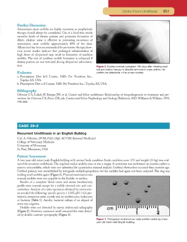
A two-year-old, intact male English bulldog with normal body condition (body condition score 3/5) and weight (24 kg) was eval-
uated for recurrent urolithiasis.The dog had voided uroliths since it was a puppy. A cystotomy was performed six months earlier to
remove urocystoliths, which were not submitted for quantitative mineral analysis. Urethral obstruction occurred three months ago.
Urethral patency was reestablished by retrograde urohydropropulsion but the uroliths had again not been analyzed. The dog was
voiding small uroliths again (Figure 1).Physical examination was
normal; uroliths were not palpable in the bladder or urethra.
Results of a complete blood count and serum biochemistry
profile were normal, except for a mildly elevated uric acid con-
centration. Analysis of a urine specimen obtained by cystocente-
sis revealed the following: specific gravity = 1.035, pH = 6.0, pro-
teinuria, numerous urate crystals and no erythrocytes, leukocytes
or bacteria (Table 1). Aerobic bacterial culture of an aliquot of
urine was negative.
Uroliths were not detected by survey abdominal radiography
(Figure 2). However, numerous small urocystoliths were detect-
ed by double-contrast cystography (Figure 3).
Figure 1. Photograph of ammonium urate uroliths voided by a two-
year-old intact male English bulldog.