
Page 838 - Small Animal Clinical Nutrition 5th Edition
P. 838
Canine Calcium Oxalate Urolithiasis 869
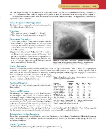
mal. Body weight was 5 kg; the dog had a normal body condition score (3/5). Survey radiographs revealed a large urinary bladder
VetBooks.ir and a radiodense urolith in the urethra at the proximal end of the os penis and several uroliths in the urinary bladder (Figure 1).
The diagnosis was urolithiasis of the lower urinary tract associated with urethral obstruction.The depression was probably a con-
sequence of postrenal azotemia.
Assess the Food and Feeding Method
The dog was fed a commercial moist adult maintenance food
twice daily and various table foods.
Questions
1. What additional assessments should be performed?
2. What should be the patient’s initial treatment plan?
Answers and Discussion
1. A blood sample should be submitted for biochemical analy-
sis to evaluate the degree of azotemia and detect concurrent
electrolyte abnormalities. A urinalysis and aerobic bacterial
culture of the urine will help predict the mineral composi-
tion of the uroliths.
2. Replacement of the patient’s fluid deficits with an appropri-
ate fluid given intravenously is important.The urinary blad-
der can be evacuated by decompressive cystocentesis to en-
hance renal elimination of waste products. Reducing pres-
sure in the urinary bladder also would facilitate retrograde Figure 1. Survey lateral radiograph of a nine-year-old male Yorkshire
urohydropropulsion of the urethrolith. terrier revealing multiple radiodense uroliths in the urinary bladder
and a radiodense urolith at the proximal end of the os penis.
Further Assessment
Results of laboratory tests revealed azotemia, hyperphosphatemia and hypobicarbonatemia (Table 1). Serum alkaline phosphatase
activity was also increased. Crystals were not observed by urine sediment examination and the urine culture was negative (Table 2).
The urethrolith was successfully flushed into the urinary bladder by retrograde urohydropropulsion. Prophylactic antimicrobials
were administered (amoxicillin-clavulanic acid, 14 mg/kg,
q12h) to prevent iatrogenic bacterial urinary tract infection Table 1. Serum biochemistry values of a nine-year-old male Yorkshire
associated with transurethral catheterization. terrier with radiodense urocystoliths.*
Reference
Additional Question Factors values Day 1 Day 2 Day 3
What is the most likely mineral composition of these radio- SUN (mg/dl) 7-28 186 141 16
Creatinine (mg/dl) 0.5-1.5 6.5 1.2 0.9
dense uroliths?
Calcium (mg/dl) 9.3-11.4 8.7 8.2 8.7
Phosphorus (mg/dl) 1.9-7.0 19.2 3.5 3.2
Answer and Discussion Sodium (mEq/l) 143-150 144 149 149
Potassium (mEq/l) 3.2-5.6 4.7 3.6 3.3
The advantages and disadvantages of medical urolith dissolu-
ALT activity (U/l) 5-62 78 ND ND
tion and surgical urolith removal can be more accurately as- Alk phos activity (U/l) 10-149 223 ND ND
sessed by predicting the mineral composition of uroliths. Mag- Total CO (mEq/l) 17-26 14.5 ND ND
2
nesium ammonium phosphate (struvite), calcium oxalate, calci-
Key: SUN = serum urea nitrogen, ALT = alanine aminotransferase,
um phosphate, silica and cystine uroliths can all be radiodense Alk phos = alkaline phosphatase, ND = not done.
(Table 3). It was surmised that this patient’s uroliths were prob- *Dietary therapy was initiated on Day 14.
ably not composed of magnesium ammonium phosphate be-
cause of the breed and gender of the dog along with findings of aciduria and a negative bacterial culture. These findings suggested
that the uroliths were not composed of struvite and therefore were not amenable to medical dissolution.
Treatment and Further Assessment
The uroliths were surgically removed following resolution of azotemia on the third day of hospitalization (Table 1). Postsurgical
radiographs verified that all uroliths were removed. Quantitative analysis revealed that the uroliths were composed of 100% calci-
um oxalate monohydrate.
Further Questions
1. Outline an appropriate feeding plan (food and feeding method) for this dog.