
Page 72 - Clinical Pearls in Cardiology
P. 72
Chapt er
3 Jugular Venous Pulse
1. What is the importance Jugular Venous Pulse?
The clinical evaluation of the Jugular Venous Pulse
(JVP) gives an idea about the performance of the right
side of the heart. The internal jugular vein is in direct
communication with the right atrium through the
superior vena cava without any intervening valves. So
the pressure changes within the right atrium are directly
reflected in the internal jugular vein. JVP is ideally
assessed on the right side of the neck.
The normal pressure in the right atrium is equivalent
to that exerted by a column of blood that is 10 to 12 cm
tall. When a person is standing or sitting upright, the
internal jugular vein is completely collapsed and when
he is lying flat, it is completely filled. When a person lies
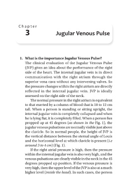
propped up at 45 degrees (as shown in the Fig. 1), the
jugular venous pulsations are normally visible just above
the clavicle. So in normal people, the height of JVP is
the vertical distance between the sternal angle of Louis
and the horizontal level at which clavicle is present (i.e
around 3 to 4 cm) (Fig. 1).
If the right atrial pressure is high, then the pressure
within the internal jugular vein is also very high, and the
venous pulsations are clearly visible in the neck in the 45
degrees propped up position. If the venous pressure is
very high, then the upper level of the JVP is also at a much
higher level (inside the head). In such cases, the person