
Page 73 - Clinical Pearls in Cardiology
P. 73
Jugular Venous Pulse 61
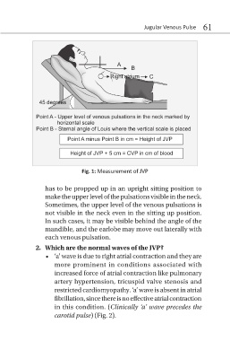
Fig. 1: Measurement of JVP
has to be propped up in an upright sitting position to
make the upper level of the pulsations visible in the neck.
Sometimes, the upper level of the venous pulsations is
not visible in the neck even in the sitting up position.
In such cases, it may be visible behind the angle of the
mandible, and the earlobe may move out laterally with
each venous pulsation.
2. Which are the normal waves of the JVP?
• ‘a’ wave is due to right atrial contraction and they are
more prominent in conditions associated with
increased force of atrial contraction like pulmonary
artery hypertension, tricuspid valve stenosis and
restricted cardiomyopathy. ‘a’ wave is absent in atrial
fibrillation, since there is no effective atrial contraction
in this condition. (Clinically ‘a’ wave precedes the
carotid pulse) (Fig. 2).