
Page 75 - Clinical Pearls in Cardiology
P. 75
Jugular Venous Pulse 63
3. What are the differences between giant ‘a’ waves and
cannon ‘a’ waves in JVP?
The giant ‘a’ waves are due to forceful right atrial
contraction towards the end of ventricular diastole
when the tricuspid valve is remaining open. It is due
to increased impedance to the right atrial emptying in
conditions like tricuspid stenosis and pulmonary artery
hypertension.
The cannon ‘a’ wave is a type of large wave that
occurs due to forceful right atrial contraction against
a closed tricuspid valve. They occur during ventricular
systole. The cannon ‘a’ waves occur when the atrial and
ventricular contractions are not coordinated (e.g.: in
complete heart block, junctional tachycardia) (Table 1).
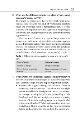
Table 1: Difference between giant ‘a’ wave and cannon ‘a’
wave
Giant ‘a’ wave Cannon ‘a’ wave
Uniform in height Variable in height
Occurs during ventricular Occurs during ventricular
diastole systole
4. What are the two important signs associated with JVP?
The two important clinical signs associated with JVP are
the Kussmaul’s sign and the hepatojugular reflux sign.
• Kussmaul’s sign: During inspiration, there is
increased venous return. This distends the right
ventricle and hence the right ventricular contraction
is stronger during inspiration (according to the
Starling’s law). Hence the right ventricular emptying
is more efficient during inspiration and there is a fall
in the upper level of JVP during inspiration in normal
individuals. But in conditions like right ventricular
failure and constrictive pericarditis, the upper level