
Page 16 - GP Fall 2020
P. 16
nosis of the tooth. After removing #26 and
allowing the extraction site to heal for two
weeks, the mesial of #27 was reevaluated
(Figure 2) and cotton roll isolation was used
while SDF was applied to the surface using
a microbrush. (Figure 3) Darkening of the
weaker dentin could be seen after less than
a minute. (Figure 4) Fluoride varnish was
applied right over the SDF. (Figure 5) Two
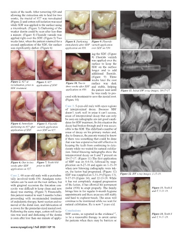
weeks later, when the patient returned for a Figure 8. Darkening Figure 9. Fluoride
second application of the SDF, the surface immediately after SDF varnish application
was significantly darker. (Figure 6) application on #30. over SDF on #30.
ing the SDF. (Figure
8) Fluoride varnish
was applied over the
surface to keep the
SDF on the surface
longer and to add
additional fluoride.
(Figure 9) Three
weeks later the root
Figure 2. #27 at Figure 3. #27 Figure 10. Two to
reevaluation, prior to application of SDF. three weeks after SDF surface was dark
SDF treatment. application on #30. and stable, helping Figure 11. Initial BW x-ray images, 10-17-17.
the patient wait until
he was ready to pro-
ceed with treatment to save the mesial root.
(Figure 10)
Case 3. 5-year-old male with open regions
of interproximal decay. Because SDF
doesn’t work well in areas it can’t access,
areas of interproximal decay that can only
be seen on radiographs are not good candi-
Figure 4. Immediate Figure 5. Fluoride dates for SDF treatment. In this situation the
darkening on #27 after varnish application decay had broken through and it was acces-
application of SDF. over SDF on #27. sible to the SDF. The child had a number of
areas of decay on his primary molars and,
due to finances, the parents wanted to know
if there was something that could be done
that was less expensive but still effective in
keeping the teeth from continuing to dete-
riorate while we waited for natural exfolia-
tion. Initial bitewing radiographs show the
interproximal decay on S and T present on
10-17-17. (Figure 11) The first application
Figure 6. One to two Figure 7. Tooth #30 of SDF was on 4-4-18, followed by reap-
weeks after SDF prior to SDF. plication on 8-27-18 and again on 1-15-19
application on #27. when new bitewing radiographs were tak-
en, the lesion had progressed. (Figure 12)
Case 2. 60-year-old male with a periodon- SDF was reapplied on 5-31-19 (Figure 13), Figure 12. BW x-ray images, 1-15-19.
tally involved tooth #30. Amalgam resto- 9-17-19 (Figure 14), and 12-13-19. While
rations can be seen on the root surface, but it has not completely stopped progression
with gingival recession the furcation con- of the lesion, it has allowed his permanent
cavity was difficult to keep clean and new molar (#30) to erupt properly. The family Figure 13. Teeth S
decay was starting. (Figure 7) Financially, brings him in for regular 6-month hygiene and T, 5-31-19.
this patient wanted to wait 5 months before appointments and these areas are still stable
proceeding with recommended treatment and hard to explorer-touch. The areas will
of endodontic therapy, hemi-section and re- continue to be monitored while we wait for
moval of the distal root, and fabrication of natural exfoliation. He is now 7 years old.
a crown for the premolar-sized mesial root.
Following the same steps, cotton roll isola- Conclusions
5-7
tion was used and darkening of the dentin SDF seems, as reported in the evidence , Figure 14. Teeth S
is seen after less than one minute of apply- to be a reasonable therapy to arrest caries and T, 9-17-19.
for patients when there may be barriers or
www.nysagd.org l Fall 2020 l GP 16