
Page 247 - The Manga Guide to Biochemistry
P. 247
The substances that pass through the ion exchange resin are collected in test tubes,
but the substances that are absorbed are separated from the resin and collected in test tubes
later by adding a liquid with a higher salt concentration. DNA polymerase a adheres to the
column and can be retrieved by adding a liquid with a salt concentration of 0.5 M (moles per
liter). (This is “sample 1.”)
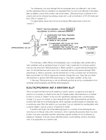
The figure below shows the method of purifying DNA polymerase α from this
“sample 1.”
“Purify” DNA polymerase α from Sample 1
according to affinity chromatography
1) Compounds that pass through
the column
2) Compounds that are absorbed
by resin in the column
Sample 1 Column Compounds that flow out with
Approximately 20 cm sodium chloride (NaCl)
DNA polymerase
Compounds that flow out with α is here!
magnesium chloride (mgCl2)
Purified!
This technique, called affinity chromatography, uses a small glass tube packed with a
resin combined with an antibody (a type of protein that’s created by the immune system)
that can only bond with DNA polymerase α. When sample 1 is passed directly through this
resin, it’s divided into two parts—one that is absorbed and one that passes through. DNA
polymerase α, which is absorbed, can be retrieved by running a solution with an extremely
high concentration (3.2 M) of magnesium chloride through the resin. Since the part that’s
retrieved here is almost 100% DNA polymerase α, it has been “purified” at this time.
In this way, DNA polymerase α can be efficiently purified by using a combination of ion
exchange chromatography and affinity chromatography.
Electrophoresis and a Western Blot
This is an experimental method for isolating a specific protein, recognizing what type of
protein is in a sample, or checking the size of a target protein. Electrophoresis is a method
in which a sample is “loaded” on top of a thin gelatinous slab (gel) and a current passes
through it to cause the sample to move through the gel. SDS-polyacrylamide gel electro-
phoresis (left side of the following figure), which separates proteins based on molecular size,
is commonly used. After the proteins are separated, they are detected by reacting them with
a special reagent, such as a dye or a fluorescent marker.
A western blot (right side of the following figure) is a method of transferring the pro-
teins that are in the gel onto a thin membrane, while retaining the positions that they were
in after they were separated, and then detecting a specific protein on the membrane by
adding an “antibody” that only reacts with that protein. Lectin blotting, which is described
next, is an application of a western blot.
Molecular Biology and the Biochemistry of Nucleic Acids 233