
Page 385 - Basic _ Clinical Pharmacology ( PDFDrive )
P. 385
CHAPTER 21 Introduction to the Pharmacology of CNS Drugs 371
and such coupling may play a role in synchronizing neuronal
discharge. However, it is unlikely that these electrical synapses are 0 Microelectrode Action A
an important site of drug action.) The events involved in synaptic enters cell potential
Axon
transmission can be summarized as follows. mV E1 Potential E1+E2 E1 E1
An action potential propagating down the axon of the presyn- Threshold
aptic neuron enters the synaptic terminal and activates voltage- –50
sensitive calcium channels in the membrane of the terminal (see –60 EPSP Resting Spatial Temporal
Figure 6–3). The calcium channels responsible for the release of –70 Potential Summation Summation
neurotransmitter are generally resistant to the calcium channel- 0
blocking agents discussed in Chapter 12 (eg, verapamil) but are B
sensitive to blockade by certain marine toxins and metal ions (see mV
Tables 21–1 and 12–4). As calcium flows into the terminal, E3 I I E3
the increase in intraterminal calcium concentration promotes –50
the fusion of synaptic vesicles with the presynaptic membrane. –60 IPSP
The neurotransmitter contained in the vesicles is released into Integration of Excitation
the synaptic cleft and diffuses to the receptors on the postsynaptic –70 and Inhibition
membrane. The neurotransmitter binds to its receptor and opens Time
channels (either directly or indirectly as described above) caus-
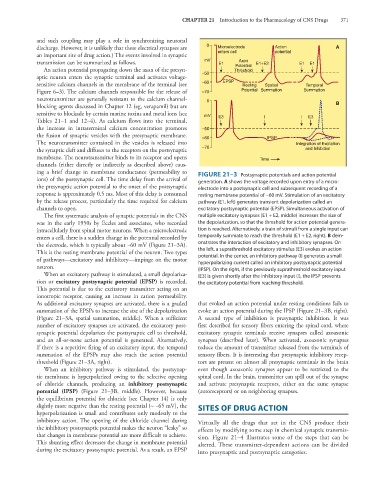
ing a brief change in membrane conductance (permeability to FIGURE 21–3 Postsynaptic potentials and action potential
ions) of the postsynaptic cell. The time delay from the arrival of generation. A shows the voltage recorded upon entry of a micro-
the presynaptic action potential to the onset of the postsynaptic electrode into a postsynaptic cell and subsequent recording of a
response is approximately 0.5 ms. Most of this delay is consumed resting membrane potential of −60 mV. Stimulation of an excitatory
by the release process, particularly the time required for calcium pathway (E1, left) generates transient depolarization called an
channels to open. excitatory postsynaptic potential (EPSP). Simultaneous activation of
The first systematic analysis of synaptic potentials in the CNS multiple excitatory synapses (E1 + E2, middle) increases the size of
was in the early 1950s by Eccles and associates, who recorded the depolarization, so that the threshold for action potential genera-
intracellularly from spinal motor neurons. When a microelectrode tion is reached. Alternatively, a train of stimuli from a single input can
enters a cell, there is a sudden change in the potential recorded by temporally summate to reach the threshold (E1 + E2, right). B dem-
the electrode, which is typically about −60 mV (Figure 21–3A). onstrates the interaction of excitatory and inhibitory synapses. On
This is the resting membrane potential of the neuron. Two types the left, a suprathreshold excitatory stimulus (E3) evokes an action
of pathways—excitatory and inhibitory—impinge on the motor potential. In the center, an inhibitory pathway (I) generates a small
hyperpolarizing current called an inhibitory postsynaptic potential
neuron. (IPSP). On the right, if the previously suprathreshold excitatory input
When an excitatory pathway is stimulated, a small depolariza- (E3) is given shortly after the inhibitory input (I), the IPSP prevents
tion or excitatory postsynaptic potential (EPSP) is recorded. the excitatory potential from reaching threshold.
This potential is due to the excitatory transmitter acting on an
ionotropic receptor, causing an increase in cation permeability.
As additional excitatory synapses are activated, there is a graded that evoked an action potential under resting conditions fails to
summation of the EPSPs to increase the size of the depolarization evoke an action potential during the IPSP (Figure 21–3B, right).
(Figure 21–3A, spatial summation, middle). When a sufficient A second type of inhibition is presynaptic inhibition. It was
number of excitatory synapses are activated, the excitatory post- first described for sensory fibers entering the spinal cord, where
synaptic potential depolarizes the postsynaptic cell to threshold, excitatory synaptic terminals receive synapses called axoaxonic
and an all-or-none action potential is generated. Alternatively, synapses (described later). When activated, axoaxonic synapses
if there is a repetitive firing of an excitatory input, the temporal reduce the amount of transmitter released from the terminals of
summation of the EPSPs may also reach the action potential sensory fibers. It is interesting that presynaptic inhibitory recep-
threshold (Figure 21–3A, right). tors are present on almost all presynaptic terminals in the brain
When an inhibitory pathway is stimulated, the postsynap- even though axoaxonic synapses appear to be restricted to the
tic membrane is hyperpolarized owing to the selective opening spinal cord. In the brain, transmitter can spill out of the synapse
of chloride channels, producing an inhibitory postsynaptic and activate presynaptic receptors, either on the same synapse
potential (IPSP) (Figure 21–3B, middle). However, because (autoreceptors) or on neighboring synapses.
the equilibrium potential for chloride (see Chapter 14) is only
slightly more negative than the resting potential (~ −65 mV), the SITES OF DRUG ACTION
hyperpolarization is small and contributes only modestly to the
inhibitory action. The opening of the chloride channel during Virtually all the drugs that act in the CNS produce their
the inhibitory postsynaptic potential makes the neuron “leaky” so effects by modifying some step in chemical synaptic transmis-
that changes in membrane potential are more difficult to achieve. sion. Figure 21–4 illustrates some of the steps that can be
This shunting effect decreases the change in membrane potential altered. These transmitter-dependent actions can be divided
during the excitatory postsynaptic potential. As a result, an EPSP into presynaptic and postsynaptic categories.