
Page 290 - parasitology for medical and clinical laboratoryprofessionals
P. 290
270 CHAPTER 12
PROCEDURE 12-4
TRICHROME STAINING PROCEDURE
Clinical Rationale
Some of the trichrome stains are modified, in that they include iron hematoxylin and
trichrome stains, called the Wheatley modification of the Gomori stain. The tech-
nique for a trichrome procedure requires less detail and is less time consuming than
the others. The stain may be made on a fresh sample or a PVA-preserved specimen.
SAF-preserved specimens do not stain as well with trichrome as with iron hematoxy-
lin. The following procedure is a standard method, but with various brands of stain,
the procedure may vary.
Stained fecal smears should be prepared from all stool specimens for posi-
tive identification of protozoan parasites in their various life stages. The Wheatley
trichrome technique is a rapid procedure that is convenient for identification of
intestinal protozoa in fresh fecal specimens. Nuclear material is easily visualized as
important tools in diagnosing particular organisms due to colors imparted to the com-
ponents of the organism. Fecal smears must be fixed using polyvinyl alcohol (PVA) or
Schaudinn solution. Modifications of the procedure and the use of other stains are
used by some to aid in observing microsporidia and acid-fast stains are often used for
Cryptosporidium, Cyclospora, and Isospora. Several different stains may be used for
parasite differentiation. Three basic varieties of the stain are used depending upon
the species of the parasite being examined (see Table 12-2).
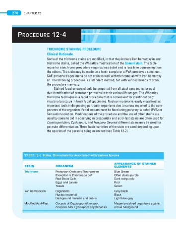
TABLE 12-2 Stains, Characteristics Associated with Various Species
APPEARANCE OF STAINED
STAIN ORGANISM ELEMENTS
Trichrome Protozoan Cysts and Trophozoites Blue Green
Exception is Entamoeba coli Often stains purple
Red Blood Cells Dark red-purple
Eggs and Larvae Red
Yeasts Green
Iron hematoxylin Organisms Gray-black
Nuclear material Black
Background material and debris Light blue-gray
Modified Acid-Fast Oocysts of Cryptosporidium spp., Magenta-stained organisms against
Isospora belli, Cyclospora cayetanensis a blue background