
Page 295 - parasitology for medical and clinical laboratoryprofessionals
P. 295
Laboratory Procedures for Identifying Parasitic Organisms and Their Ova 275
Source: Centers for Disease Control and Prevention (CDC)
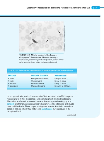
FIGURE 12-8 Malarial parasites in blood smears.
Micrograph of Giemsa-stained thin smear showing a
Plasmodium falciparum gametocyte (bottom, double arrow)
and several ring forms within erythrocytes (arrows).
TABLE 12-3 Fever cycles characteristic of malaria species that infect humans
SPECIES DISEASE CAUSED PAROXYSMS
P. vivax Benign tertian malaria Every 48 hours
P. ovale Ovale malaria Every 48 hours
P. malariae Quartan malaria Every 72 hours
P. falciparum Malignant malaria Every 36 to 38 hours
recurs periodically), each of the merozoite-filled red blood cells (RBCs) rupture
releasing 12 to 24 free merozoit es and malarial pigment into the blood stream.
Merozoites are formed by asexual reproduction through the breaking up of a
schizont (another stage in asexual reproduction of some protozoans) and invade
other red blood cells. These stages are ingested into the Anopheles mosquito in
cases of malaria, where they mature into gametocytes that reproduce in the
mosquito’s blood.
(continues)