
Page 299 - parasitology for medical and clinical laboratoryprofessionals
P. 299
Laboratory Procedures for Identifying Parasitic Organisms and Their Ova 279
3. Allow the water to lyse the RBCs, which usually takes approximately 3 minutes, and
remove excess water from slide. Allow slide to air-dry com pletely before staining.
If available, a positive control slide should be stained with all malaria specimens.
4. Place the slide in a Coplin jar containing Wright-Giemsa stain (Quick-Stain) for
10 seconds.
5. Place slide in a Coplin jar of distilled water for 20 seconds or more to achieve a
desired color balance.
6. Drain the slide and allow to air-dry in an upright position, by allowing it to stand
on its end.
Examining the Stained Slides
1. Examine the stained slide under a microscope using the oil immersion
objective (1000 3).
2. The various stages of blood and tissue parasites are often similar for different
species. Determining the various stages (Table 12-5) of the life cycle for these
organisms is invaluable in providing a diagnosis.
3. Examine thick smears thoroughly for the presence of malarial parasites.
Remember that the cytoplasm of the Plasmodium species stains robin egg
blue and the nuclear chromatin stains crimson or violet. Examine at least
100 oil immersion fields on each thick film.
4. The thin films are used primarily for speciating the Plasmo dium organism. How-
ever, even if no parasites are discovered on the thick films, the thin films must still
be examined. View at least 200 fields on each thin smear before reporting a nega-
tive result.
5. When malarial parasites are observed, identify the organism using the diagram
(see Figure 12-12).
6. The results obtained from all positive malarial slide preparations should be
phoned to the attend ing physician or his or her designate as soon as possible.
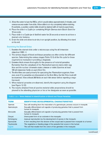
TABLE 12-5 Terms Related to Identification of Blood and Tissue Parasites
TERM IDENTIFYING DEVELOPMENTAL CHARACTERISTICS
Gamete Sex cell resulting from the maturation of a gametocyte; process occurs in mosquito
Gametocyte Sexually differentiated cell capable of producing gametes that is passed from
human to mosquito
Merozoite Sexually differentiated cell capable of producing gametes that is passed from
human to mosquito
Oocyst Unencysted form of an ookinete in the mosquito
Schizogony Asexual reproduction by the development of spores in the mosquito
Sporozoite A form resulting from the division of the oocyst and is passed from mosquito to a human
Trophozoite The vegetative or feeding stage of the parasite that occurs in the human
Zygote The cell that results from the union of two gametes in the mosquito
(continues)