
Page 302 - parasitology for medical and clinical laboratoryprofessionals
P. 302
282 CHAPTER 12
Source: Centers for Disease Control and Prevention (CDC)
Delmar/Cengage Learning
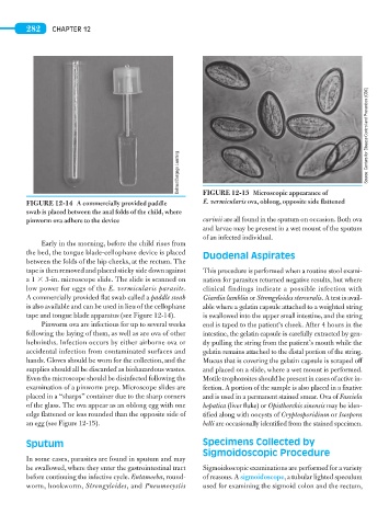
E. vermicularis ova, oblong, opposite side flattened
FIGURE 12-14 A commercially provided paddle FIGURE 12-15 Microscopic appearance of
swab is placed between the anal folds of the child, where
pinworm ova adhere to the device carinii are all found in the sputum on occasion. Both ova
and larvae may be present in a wet mount of the sputum
of an infected individual.
Early in the morning, before the child rises from
the bed, the tongue blade-cellophane device is placed Duodenal Aspirates
between the folds of the hip cheeks, at the rectum. The
tape is then removed and placed sticky side down against This procedure is performed when a routine stool exami-
a 1 3 3-in. microscope slide. The slide is scanned on nation for parasites returned negative results, but where
low power for eggs of the E. vermicularis parasite. clinical findings indicate a possible infection with
A commercially provided flat swab called a paddle swab Giardia lamblia or Strongyloides stercoralis. A test is avail-
is also available and can be used in lieu of the cellophane able where a gelatin capsule attached to a weighted string
tape and tongue blade apparatus (see Figure 12-14). is swallowed into the upper small intestine, and the string
Pinworm ova are infectious for up to several weeks end is taped to the patient’s cheek. After 4 hours in the
following the laying of them, as well as are ova of other intestine, the gelatin capsule is carefully extracted by gen-
helminths. Infection occurs by either airborne ova or tly pulling the string from the patient’s mouth while the
accidental infection from contaminated surfaces and gelatin remains attached to the distal portion of the string.
hands. Gloves should be worn for the collection, and the Mucus that is covering the gelatin capsule is scraped off
supplies should all be discarded as biohazardous wastes. and placed on a slide, where a wet mount is performed.
Even the microscope should be disinfected following the Motile trophozoites should be present in cases of active in-
examination of a pinworm prep. Microscope slides are fection. A portion of the sample is also placed in a fixative
placed in a “sharps” container due to the sharp corners and is used in a permanent stained smear. Ova of Fasciola
of the glass. The ova appear as an oblong egg with one hepatica (liver fluke) or Opisthorchis sinensis may be iden-
edge flattened or less rounded than the opposite side of tified along with oocysts of Cryptosporidium or Isospora
an egg (see Figure 12-15). belli are occasionally identified from the stained specimen.
Sputum Specimens Collected by
Sigmoidoscopic Procedure
In some cases, parasites are found in sputum and may
be swallowed, where they enter the gastrointestinal tract Sigmoidoscopic examinations are performed for a variety
before continuing the infective cycle. Entamoeba, round- of reasons. A sigmoidoscope, a tubular lighted speculum
worm, hookworm, Strongyloides, and Pneumocystis used for examining the sigmoid colon and the rectum,