
Page 156 - Atlas of Histology with Functional Correlations
P. 156
islet. B. High magnification of the endocrine and exocrine pancreas. Stain:
hematoxylin and eosin. High magnification.
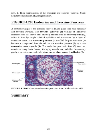
FIGURE 4.20 | Endocrine and Exocrine Pancreas
A photomicrograph of the pancreas shows a mixed gland with both endocrine
and exocrine portions. The exocrine pancreas (3) consists of numerous
secretory acini that deliver their secretory material into the excretory duct (1),
which is lined by simple cuboidal epithelium and surrounded by a layer of
connective tissue. The endocrine pancreas (5) is called the pancreatic islet (5)
because it is separated from the cells of the exocrine pancreas (3) by a thin
connective tissue capsule (4). The endocrine pancreatic islet (5) does not
contain excretory ducts. Instead, it is highly vascularized, and all of the secretory
products leave the pancreatic islet via numerous blood vessels (capillaries) (2).
FIGURE 4.20 ■ Endocrine and exocrine pancreas. Stain: Mallory-Azan. ×100.
Summary
155