
Page 154 - Atlas of Histology with Functional Correlations
P. 154
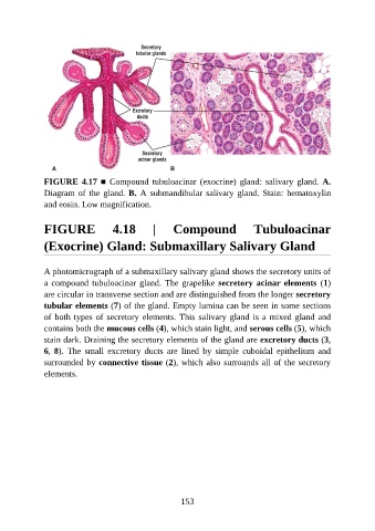
FIGURE 4.17 ■  Compound  tubuloacinar  (exocrine)  gland:  salivary  gland.  A.

               Diagram of the gland. B. A submandibular salivary gland. Stain: hematoxylin
               and eosin. Low magnification.


               FIGURE                4.18         |     Compound                  Tubuloacinar


               (Exocrine) Gland: Submaxillary Salivary Gland


               A photomicrograph of a submaxillary salivary gland shows the secretory units of
               a compound tubuloacinar gland. The grapelike secretory acinar elements  (1)

               are circular in transverse section and are distinguished from the longer secretory
               tubular elements (7) of the gland. Empty lumina can be seen in some sections
               of both types of secretory elements. This salivary gland is a mixed gland and
               contains both the mucous cells (4), which stain light, and serous cells (5), which

               stain dark. Draining the secretory elements of the gland are excretory ducts (3,
               6, 8).  The  small  excretory  ducts  are  lined  by  simple  cuboidal  epithelium  and
               surrounded by connective tissue (2), which also surrounds all of the secretory
               elements.


                                                          153