
Page 226 - Atlas of Histology with Functional Correlations
P. 226
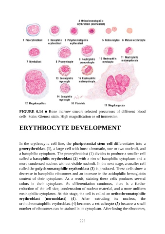
FIGURE  6.14  ■  Bone  marrow  smear:  selected  precursors  of  different  blood
               cells. Stain: Giemsa stain. High magnification or oil immersion.


               ERYTHROCYTE DEVELOPMENT


               In  the  erythrocytic  cell  line,  the  pluripotential  stem  cell  differentiates  into  a
               proerythroblast (1), a large cell with loose chromatin, one or two nucleoli, and
               a basophilic cytoplasm. The proerythroblast (1) divides to produce a smaller cell

               called a basophilic erythroblast (2) with a rim of basophilic cytoplasm and a
               more condensed nucleus without visible nucleoli. In the next stage, a smaller cell
               called the polychromatophilic erythroblast (3) is produced. These cells show a
               decrease in basophilic ribosomes and an increase in the acidophilic hemoglobin

               content  of  their  cytoplasm.  As  a  result,  staining  these  cells  produces  several
               colors  in  their  cytoplasm.  As  differentiation  continues,  there  is  a  further
               reduction of the cell size, condensation of nuclear material, and a more uniform
               eosinophilic cytoplasm. At this stage, the cell is called an orthochromatophilic

               erythroblast  (normoblast)  (4).             After    extruding      its   nucleus,     the
               orthochromatophilic erythroblast (4) becomes a reticulocyte (5) because a small
               number of ribosomes can be stained in its cytoplasm. After losing the ribosomes,


                                                          225