
Page 225 - Atlas of Histology with Functional Correlations
P. 225
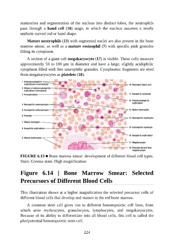
maturation and segmentation of the nucleus into distinct lobes, the neutrophils

               pass  through  a  band  cell  (10)  stage,  in  which  the  nucleus  assumes  a  nearly
               uniform curved rod or band shape.

                   Mature neutrophils (13) with segmented nuclei are also present in the bone
               marrow smear, as well as a mature eosinophil (7) with specific pink granules
               filling its cytoplasm.

                   A section of a giant cell megakaryocyte (17) is visible. These cells measure

               approximately 50 to 100 μm in diameter and have a large, slightly acidophilic
               cytoplasm filled with fine azurophilic granules. Cytoplasmic fragments are shed
               from megakaryocytes as platelets (18).


               FIGURE 6.13 ■ Bone marrow smear: development of different blood cell types.
               Stain: Giemsa stain. High magnification.


               Figure  6.14  |  Bone  Marrow  Smear:  Selected

               Precursors of Different Blood Cells


               This illustration shows at a higher magnification the selected precursor cells of
               different blood cells that develop and mature in the red bone marrow.

                   A common stem cell gives rise to different hematopoietic cell lines, from
               which  arise  erythrocytes,  granulocytes,  lymphocytes,  and  megakaryocytes.

               Because of its ability to differentiate into all blood cells, this cell is called the
               pluripotential hematopoietic stem cell.


                                                          224