
Page 223 - Atlas of Histology with Functional Correlations
P. 223
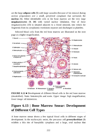
are the large adipose cells (5) with large vacuoles (because of fat removal during

               section  preparation)  and  a  small,  peripheral  cytoplasm  that  surrounds  the
               nucleus  (5).  Other  identifiable  cells  in  the  bone  marrow  are  the  very  large
               megakaryocytes  (9,  10)  with  varied  nuclear  lobulation.  One  of  these
               megakaryocytes  (10)  is  situated  adjacent  to  a  blood  sinusoid,  into  which  the
               fragments from its cytoplasmic extensions separate and discharged as platelets.


                   Selected blood cells from the red bone marrow are illustrated on the next
               page at a higher magnification.


               FIGURE 6.12 ■ Development of different blood cells in the red bone marrow

               (decalcified). Stain:  hematoxylin  and  eosin.  Upper image:  high  magnification;
               lower image: oil immersion.


               Figure  6.13  |  Bone  Marrow  Smear:  Development

               of Different Cell Types


               A  bone  marrow  smear  shows  a  few  typical  blood  cells  in  different  stages  of
               development. In the erythrocytic series, the precursor cell proerythroblast (3)

               exhibits  a  thin  rim  of  basophilic  cytoplasm  and  a  large,  oval  nucleus  that


                                                          222