
Page 256 - Atlas of Histology with Functional Correlations
P. 256
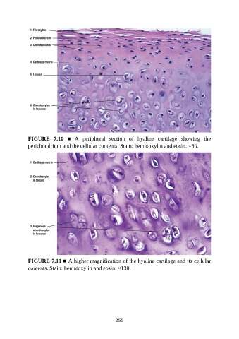
FIGURE  7.10  ■  A  peripheral  section  of  hyaline  cartilage  showing  the

               perichondrium and the cellular contents. Stain: hematoxylin and eosin. ×80.


               FIGURE 7.11 ■ A higher magnification of the hyaline cartilage and its cellular

               contents. Stain: hematoxylin and eosin. ×130.


                                                          255