
Page 504 - Atlas of Histology with Functional Correlations
P. 504
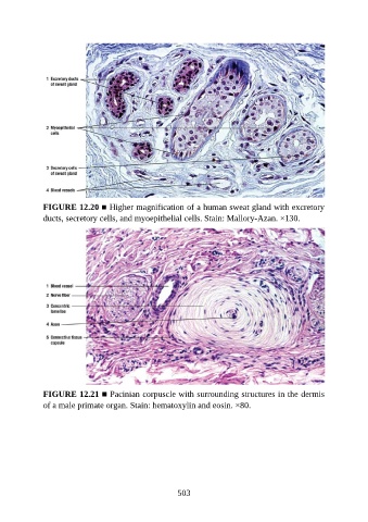
FIGURE 12.20 ■ Higher magnification of a human sweat gland with excretory
               ducts, secretory cells, and myoepithelial cells. Stain: Mallory-Azan. ×130.


               FIGURE 12.21 ■ Pacinian corpuscle with surrounding structures in the dermis
               of a male primate organ. Stain: hematoxylin and eosin. ×80.


                                                          503